1FJT

THERMOLYSIN (50% ACETONITRILE SOAKED CRYSTALS)


Experimental Data Snapshot

  • Method: X-RAY DIFFRACTION
  • Resolution: 2.20 Å
  • R-Value Free: 0.209 
  • R-Value Work: 0.149 

wwPDB Validation   3D Report Full Report


This is version 1.3 of the entry. See complete history


Literature

Experimental and computational mapping of the binding surface of a crystalline protein.

English, A.C.Groom, C.R.Hubbard, R.E.

(2001) Protein Eng 14: 47-59

  • DOI: https://doi.org/10.1093/protein/14.1.47
  • Primary Citation of Related Structures:  
    1FJ3, 1FJO, 1FJQ, 1FJT, 1FJU, 1FJV, 1FJW

  • PubMed Abstract: 

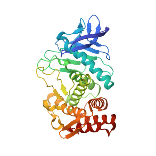
    Multiple Solvent Crystal Structures (MSCS) is a crystallographic technique to identify energetically favorable positions and orientations of small organic molecules on the surface of proteins. We determined the high-resolution crystal structures of thermolysin (TLN), generated from crystals soaked in 50--70% acetone, 50--80% acetonitrile and 50 mM phenol. The structures of the protein in the aqueous-organic mixtures are essentially the same as the native enzyme and a number of solvent interaction sites were identified. The distribution of probe molecules shows clusters in the main specificity pocket of the active site and a buried subsite. Within the active site, we compared the experimentally determined solvent positions with predictions from two computational functional group mapping techniques, GRID and Multiple Copy Simultaneous Search (MCSS). The experimentally determined small molecule positions are consistent with the structures of known protein--ligand complexes of TLN.


  • Organizational Affiliation

    Structural Biology Laboratory, Department of Chemistry, University of York, Heslington, York YO10 5DD, UK.


Macromolecules
Find similar proteins by:  (by identity cutoff)  |  3D Structure
Entity ID: 1
MoleculeChains Sequence LengthOrganismDetailsImage
THERMOLYSIN316Bacillus thermoproteolyticusMutation(s): 0 
EC: 3.4.24.27
UniProt
Find proteins for P00800 (Bacillus thermoproteolyticus)
Explore P00800 
Go to UniProtKB:  P00800
Entity Groups  
Sequence Clusters30% Identity50% Identity70% Identity90% Identity95% Identity100% Identity
UniProt GroupP00800
Sequence Annotations
Expand
  • Reference Sequence
Small Molecules
Ligands 5 Unique
IDChains Name / Formula / InChI Key2D Diagram3D Interactions
LYS
Query on LYS

Download Ideal Coordinates CCD File 
I [auth A]LYSINE
C6 H15 N2 O2
KDXKERNSBIXSRK-YFKPBYRVSA-O
VAL
Query on VAL

Download Ideal Coordinates CCD File 
H [auth A]VALINE
C5 H11 N O2
KZSNJWFQEVHDMF-BYPYZUCNSA-N
DMS
Query on DMS

Download Ideal Coordinates CCD File 
G [auth A]DIMETHYL SULFOXIDE
C2 H6 O S
IAZDPXIOMUYVGZ-UHFFFAOYSA-N
ZN
Query on ZN

Download Ideal Coordinates CCD File 
B [auth A]ZINC ION
Zn
PTFCDOFLOPIGGS-UHFFFAOYSA-N
CA
Query on CA

Download Ideal Coordinates CCD File 
C [auth A],
D [auth A],
E [auth A],
F [auth A]
CALCIUM ION
Ca
BHPQYMZQTOCNFJ-UHFFFAOYSA-N
Experimental Data & Validation

Experimental Data

  • Method: X-RAY DIFFRACTION
  • Resolution: 2.20 Å
  • R-Value Free: 0.209 
  • R-Value Work: 0.149 
  • Space Group: P 61 2 2
Unit Cell:
Length ( Å )Angle ( ˚ )
a = 93.811α = 90
b = 93.811β = 90
c = 131.101γ = 120
Software Package:
Software NamePurpose
REFMACrefinement
DENZOdata reduction
CCP4data scaling

Structure Validation

View Full Validation Report


Entry History 

Deposition Data

Revision History  (Full details and data files)

  • Version 1.0: 2001-04-18
    Type: Initial release
  • Version 1.1: 2008-04-27
    Changes: Version format compliance
  • Version 1.2: 2011-07-13
    Changes: Version format compliance
  • Version 1.3: 2024-02-07
    Changes: Data collection, Database references, Derived calculations