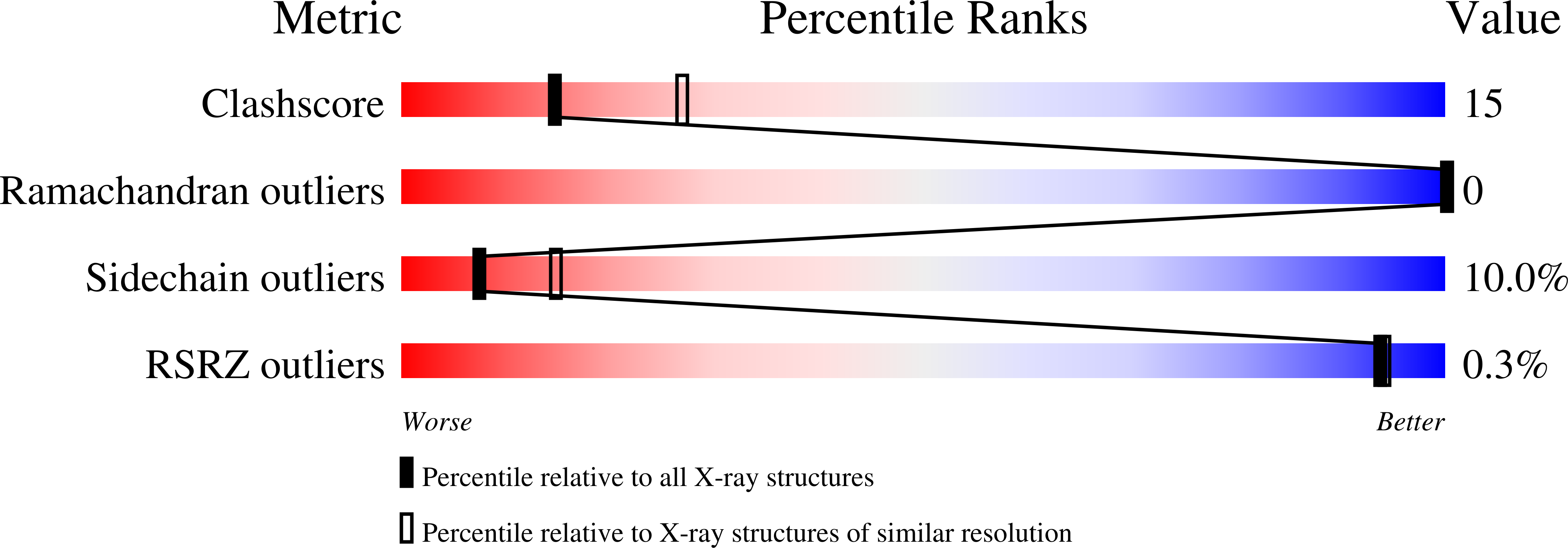
Buried charged surface in proteins.
Kajander, T., Kahn, P.C., Passila, S.H., Cohen, D.C., Lehtio, L., Adolfsen, W., Warwicker, J., Schell, U., Goldman, A.(2000) Structure 8: 1203-1214
- PubMed: 11080642
- DOI: https://doi.org/10.1016/s0969-2126(00)00520-7
- Primary Citation of Related Structures:
1F9C - PubMed Abstract:
The traditional picture of charged amino acids in globular proteins is that they are almost exclusively on the outside exposed to the solvent. Buried charges, when they do occur, are assumed to play an essential role in catalysis and ligand binding, or in stabilizing structure as, for instance, helix caps. By analyzing the amount and distribution of buried charged surface and charges in proteins over a broad range of protein sizes, we show that buried charge is much more common than is generally believed. We also show that the amount of buried charge rises with protein size in a manner which differs from other types of surfaces, especially aromatic and polar uncharged surfaces. In large proteins such as hemocyanin, 35% of all charges are greater than 75% buried. Furthermore, at all sizes few charged groups are fully exposed. As an experimental test, we show that replacement of the buried D178 of muconate lactonizing enzyme by N stabilizes the enzyme by 4.2 degrees C without any change in crystallographic structure. In addition, free energy calculations of stability support the experimental results. Nature may use charge burial to reduce protein stability; not all buried charges are fully stabilized by a prearranged protein environment. Consistent with this view, thermophilic proteins often have less buried charge. Modifying the amount of buried charge at carefully chosen sites may thus provide a general route for changing the thermophilicity or psychrophilicity of proteins.
Organizational Affiliation:
Centre for Biotechnology, University of Turku, Turku, Finland.


