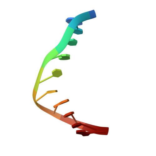
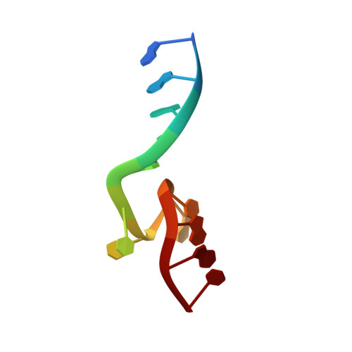
Hybrid-hybrid matrix structural refinement of a DNA three-way junction from 3D NOESY-NOESY.
Thiviyanathan, V., Luxon, B.A., Leontis, N.B., Illangasekare, N., Donne, D.G., Gorenstein, D.G.(1999) J Biomol NMR 14: 209-221
- PubMed: 10481274
- DOI: https://doi.org/10.1023/a:1008330011425
- Primary Citation of Related Structures:
1EKW - PubMed Abstract:
Homonuclear 3D NOESY-NOESY has shown great promise for the structural refinement of large biomolecules. A computationally efficient hybrid-hybrid relaxation matrix refinement methodology, using 3D NOESY-NOESY data, was used to refine the structure of a DNA three-way junction having two unpaired bases at the branch point of the junction. The NMR data and the relaxation matrix refinement confirm that the DNA three-way junction exists in a folded conformation with two of the helical stems stacked upon each other. The third unstacked stem extends away from the junction, forming an acute angle (approximately 60 degrees) with the stacked stems. The two unpaired bases are stacked upon each other and are exposed to the solvent. Helical parameters for the bases in all three strands show slight deviations from typical values expected for right-handed B-form DNA. Inter-nucleotide imino-imino NOEs between the bases at the branch point of the junction show that the junction region is well defined. The helical stems show mobility (+/- 20 degrees) indicating dynamic processes around the junction region. The unstacked helical stem adjacent to the unpaired bases shows greater mobility compared to the other two stems. The results from this study indicate that the 3D hybrid-hybrid matrix MORASS refinement methodology, by combining the spectral dispersion of 3D NOESY-NOESY and the computational efficiency of 2D refinement programs, provides an accurate and robust means for structure determination of large biomolecules. Our results also indicate that the 3D MORASS method gives higher quality structures compared to the 2D complete relaxation matrix refinement method.
Organizational Affiliation:
Sealy Center for Structural Biology, University of Texas Medical Branch, Galveston 77555-1157, USA.