1EHV

A NEW CRYSTAL STRUCTURE FOR THE DODECAMER C-G-C-G-A-A-T-T-C-G-C-G: SYMMETRY EFFECTS ON SEQUENCE-DEPENDENT DNA STRUCTURE

  • Classification: DNA
  • Mutation(s): No 

  • Deposited: 2000-02-23 Released: 2000-07-14 
  • Deposition Author(s): Johansson, E.M.

Experimental Data Snapshot

  • Method: X-RAY DIFFRACTION
  • Resolution: 1.80 Å
  • R-Value Free: 0.312 
  • R-Value Work: 0.238 
  • R-Value Observed: 0.224 

wwPDB Validation   3D Report Full Report


This is version 1.4 of the entry. See complete history


Literature

A new crystal form for the dodecamer C-G-C-G-A-A-T-T-C-G-C-G: symmetry effects on sequence-dependent DNA structure.

Johansson, E.Parkinson, G.Neidle, S.

(2000) J Mol Biol 300: 551-561

  • DOI: https://doi.org/10.1006/jmbi.2000.3907
  • Primary Citation of Related Structures:  
    1EHV

  • PubMed Abstract: 

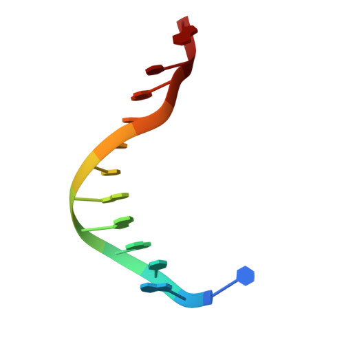
    The dodecanucleotide d(CGCGAATTCGCG) has been crystallised in the space group P3(2)12, representing a new crystal form for this sequence. The structure has been solved by molecular replacement and refined at 1.8 A resolution. The present structure contrasts with previous ones for this sequence since it is situated on a crystallographic 2-fold axis, and the crystal symmetry reflects the palindromic nature of this sequence. Some features accord with previous observations, notably that the minor groove is hydrated with a continuous spine of solvent. There is no evidence of alkali metal ions within this spine. The minor groove retains its narrow width, although it is now symmetric and extends over the A/T tract. Various base and base-pair morphological parameters have been examined. Their values do not show significant correlations with earlier reports, suggesting that crystal packing effects on them are more dominant than has been hitherto realised.


  • Organizational Affiliation

    Institute of Cancer Research, CRC Biomolecular Structure Unit, Chester Beatty Laboratories, Fulham Road, London, SW3 6JB, UK.


Macromolecules

Find similar nucleic acids by:  Sequence   |   3D Structure  

Entity ID: 1
MoleculeChains LengthOrganismImage
DNA (5'-D(*CP*GP*CP*GP*AP*AP*TP*TP*CP*GP*CP*G)-3')12N/A
Sequence Annotations
Expand
  • Reference Sequence
Experimental Data & Validation

Experimental Data

  • Method: X-RAY DIFFRACTION
  • Resolution: 1.80 Å
  • R-Value Free: 0.312 
  • R-Value Work: 0.238 
  • R-Value Observed: 0.224 
  • Space Group: P 32 1 2
Unit Cell:
Length ( Å )Angle ( ˚ )
a = 26.39α = 90
b = 26.39β = 90
c = 98.78γ = 120
Software Package:
Software NamePurpose
X-PLORmodel building
SHELXL-97refinement
MAR345data collection
SCALEPACKdata scaling
X-PLORphasing

Structure Validation

View Full Validation Report


Entry History 

Deposition Data

Revision History  (Full details and data files)

  • Version 1.0: 2000-07-14
    Type: Initial release
  • Version 1.1: 2008-04-27
    Changes: Version format compliance
  • Version 1.2: 2011-07-13
    Changes: Version format compliance
  • Version 1.3: 2017-10-04
    Changes: Refinement description
  • Version 1.4: 2024-02-07
    Changes: Data collection, Database references