Solution Structure and Dynamics of the Central Ccp Module Pair of a Poxvirus Complement Control Protein
Henderson, C.E., Bromek, K., Mullin, N.P., Smith, B.O., Uhrin, D., Barlow, P.N.(2001) J Mol Biol 307: 323
- PubMed: 11243823
- DOI: https://doi.org/10.1006/jmbi.2000.4477
- Primary Citation of Related Structures:
1E5G - PubMed Abstract:
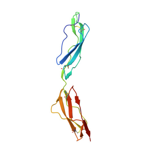
The complement control protein (CCP) module (also known as SCR, CCP or sushi domain) is prevalent amongst proteins that regulate complement activation. Functional and mutagenesis studies have shown that in most cases two or more neighbouring CCP modules form specific binding sites for other molecules. Hence the orientation in space of a CCP module with respect to its neighbours and the flexibility of the intermodular junction are likely to be critical for function. Vaccinia virus complement control protein (VCP) is a complement regulatory protein composed of four tandemly arranged CCP modules. The solution structure of the carboxy-terminal half of this protein (CCP modules 3 and 4) has been solved previously. The structure of the central portion (modules 2 and 3, VCP approximately 2,3) has now also been solved using NMR spectroscopy at 37 degrees C. In addition, the backbone dynamics of VCP approximately 2,3 have been characterised by analysis of its (15)N relaxation parameters. Module 2 has a typical CCP module structure while module 3 in the context of VCP approximately 2,3 has some modest but significant differences in structure and dynamics to module 3 within the 3,4 pair. Modules 2 and 3 do not share an extensive interface, unlike modules 3 and 4. Only two possible NOEs were identified between the bodies of the modules, but a total of 40 NOEs between the short intermodular linker of VCP approximately 2,3 and the bodies of the two modules determines a preferred, elongated, orientation of the two modules in the calculated structures. The anisotropy of rotational diffusion has been characterised from (15)N relaxation data, and this indicates that the time-averaged structure is more compact than suggested by (1)H-(1)H NOEs. The data are consistent with the presence of many intermodular orientations, some of which are kinked, undergoing interconversion on a 10(-8)-10(-6) second time-scale. A reconstructed representation of modules 2-4 allows visualisation of the spatial arrangement of the 11 substitutions that occur in the more potent complement inhibitor from Variola (small pox) virus.
Organizational Affiliation:
The Edinburgh Centre for Protein Technology, the University of Edinburgh, the Joseph Black Chemistry Building, the King's Buildings, West Mains Road, Edinburgh EH9 3JJ, UK.