NMR solution structure of the theta subunit of DNA polymerase III from Escherichia coli.
Keniry, M.A., Berthon, H.A., Yang, J.Y., Miles, C.S., Dixon, N.E.(2000) Protein Sci 9: 721-723
- PubMed: 10794414
- DOI: https://doi.org/10.1110/ps.9.4.721
- Primary Citation of Related Structures:
1DU2 - PubMed Abstract:
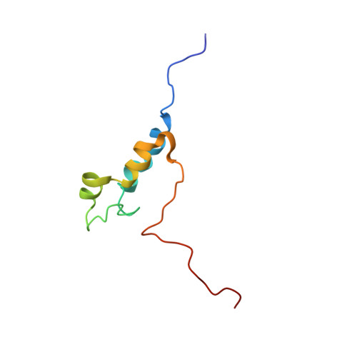
The catalytic core of Escherichia coli DNA polymerase III contains three tightly associated subunits (alpha, epsilon, and theta). The theta subunit is the smallest, but the least understood of the three. As a first step in a program aimed at understanding its function, the structure of the theta subunit has been determined by triple-resonance multidimensional NMR spectroscopy. Although only a small protein, theta was difficult to assign fully because approximately one-third of the protein is unstructured, and some sections of the remaining structured parts undergo intermediate intramolecular exchange. The secondary structure was deduced from the characteristic nuclear Overhauser effect patterns, the 3J(HN alpha) coupling constants and the consensus chemical shift index. The C-terminal third of the protein, which has many charged and hydrophilic amino acid residues, has no well-defined secondary structure and exists in a highly dynamic state. The N-terminal two-thirds has three helical segments (Gln10-Asp19, Glu38-Glu43, and His47-Glu54), one short extended segment (Pro34-Ala37), and a long loop (Ala20-Glu29), of which part may undergo intermediate conformational exchange. Solution of the three-dimensional structure by NMR techniques revealed that the helices fold in such a way that the surface of theta is bipolar, with one face of the protein containing most of the acidic residues and the other face containing most of the long chain basic residues. Preliminary chemical shift mapping experiments with a domain of the epsilon subunit have identified a loop region (Ala20-Glu29) in theta as the site of association with epsilon.
Organizational Affiliation:
Research School of Chemistry, The Australian National University, Canberra, ACT. max@rsc.anu.edu.au