1DT7

SOLUTION STRUCTURE OF THE C-TERMINAL NEGATIVE REGULATORY DOMAIN OF P53 IN A COMPLEX WITH CA2+-BOUND S100B(BB)


Experimental Data Snapshot

  • Method: SOLUTION NMR
  • Conformers Calculated: 40 
  • Conformers Submitted: 40 
  • Selection Criteria: structure with the lowest energy and the least restraint violations 

wwPDB Validation   3D Report Full Report


This is version 1.3 of the entry. See complete history


Literature

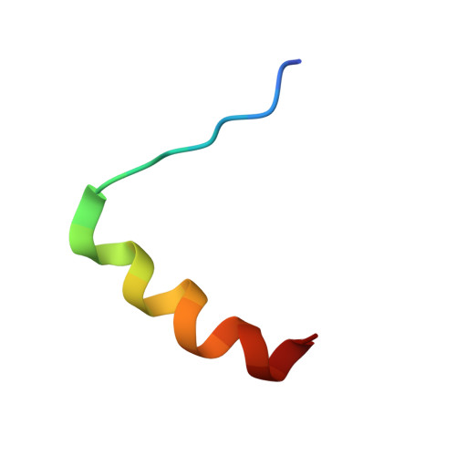
Structure of the negative regulatory domain of p53 bound to S100B(betabeta).

Rustandi, R.R.Baldisseri, D.M.Weber, D.J.

(2000) Nat Struct Biol 7: 570-574

  • DOI: https://doi.org/10.1038/76797
  • Primary Citation of Related Structures:  
    1DT7

  • PubMed Abstract: 

    A Ca2+ dependent conformational change in dimeric S100B(betabeta) is required for it to bind p53 and inhibit phosphorylation of this tumor suppressor in its C-terminal negative regulatory domain. A peptide derived from this region of p53 (residues 367-388) was found to have no regular structure in its native form by NMR spectroscopy, but becomes helical when bound to Ca2+ loaded S100B(betabeta). The three-dimensional structure of this complex reveals several favorable hydrophobic and electrostatic interactions between S100B(betabeta) and the p53 peptide in the binding pocket, where S100B(betabeta) sterically blocks sites of phosphorylation and acetylation on p53 that are important for transcription activation.


  • Organizational Affiliation

    Department of Biochemistry and Molecular Biology, University of Maryland School of Medicine, 108 N. Greene Street, Baltimore, Maryland 21201, USA.


Macromolecules
Find similar proteins by:  (by identity cutoff)  |  3D Structure
Entity ID: 1
MoleculeChains Sequence LengthOrganismDetailsImage
S100 CALCIUM-BINDING PROTEIN
A, B
92Rattus norvegicusMutation(s): 0 
UniProt
Find proteins for P04631 (Rattus norvegicus)
Explore P04631 
Go to UniProtKB:  P04631
Entity Groups  
Sequence Clusters30% Identity50% Identity70% Identity90% Identity95% Identity100% Identity
UniProt GroupP04631
Sequence Annotations
Expand
  • Reference Sequence

Find similar proteins by:  Sequence   |   3D Structure  

Entity ID: 2
MoleculeChains Sequence LengthOrganismDetailsImage
CELLULAR TUMOR ANTIGEN P53C [auth X],
D [auth Y]
22N/AMutation(s): 0 
UniProt & NIH Common Fund Data Resources
Find proteins for P04637-7 (Homo sapiens)
Explore P04637-7 
Go to UniProtKB:  P04637-7
GTEx:  ENSG00000141510 
Entity Groups  
Sequence Clusters30% Identity50% Identity70% Identity90% Identity95% Identity100% Identity
UniProt GroupP04637-7
Sequence Annotations
Expand
  • Reference Sequence
Experimental Data & Validation

Experimental Data

  • Method: SOLUTION NMR
  • Conformers Calculated: 40 
  • Conformers Submitted: 40 
  • Selection Criteria: structure with the lowest energy and the least restraint violations 

Structure Validation

View Full Validation Report


Entry History 

Deposition Data

Revision History  (Full details and data files)

  • Version 1.0: 2000-07-26
    Type: Initial release
  • Version 1.1: 2008-04-27
    Changes: Version format compliance
  • Version 1.2: 2011-07-13
    Changes: Version format compliance
  • Version 1.3: 2016-04-27
    Changes: Other