1BFN

BETA-AMYLASE/BETA-CYCLODEXTRIN COMPLEX


Experimental Data Snapshot

  • Method: X-RAY DIFFRACTION
  • Resolution: 2.07 Å
  • R-Value Free: 0.211 
  • R-Value Work: 0.158 
  • R-Value Observed: 0.158 

wwPDB Validation   3D Report Full Report


This is version 2.1 of the entry. See complete history


Literature

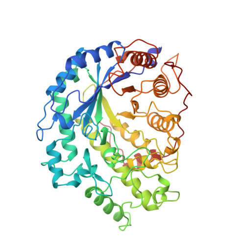
Crystal structure of recombinant soybean beta-amylase complexed with beta-cyclodextrin.

Adachi, M.Mikami, B.Katsube, T.Utsumi, S.

(1998) J Biol Chem 273: 19859-19865

  • DOI: https://doi.org/10.1074/jbc.273.31.19859
  • Primary Citation of Related Structures:  
    1BFN

  • PubMed Abstract: 

    In order to study the interaction of soybean beta-amylase with substrate, we solved the crystal structure of beta-cyclodextrin-enzyme complex and compared it with that of alpha-cyclodextrin-enzyme complex. The enzyme was expressed in Escherichia coli at a high level as a soluble and catalytically active protein. The purified recombinant enzyme had properties nearly identical to those of native soybean beta-amylase and formed the same crystals as the native enzyme. The crystal structure of recombinant enzyme complexed with beta-cyclodextrin was refined at 2. 07-A resolution with a final crystallographic R value of 15.8% (Rfree = 21.1%). The root mean square deviation in the position of C-alpha atoms between this recombinant enzyme and the native enzyme was 0.22 A. These results indicate that the expression system established here is suitable for studying structure-function relationships of beta-amylase. The conformation of the bound beta-cyclodextrin takes an ellipsoid shape in contrast to the circular shape of the bound alpha-cyclodextrin. The cyclodextrins shared mainly two glucose binding sites, 3 and 4. The glucose residue 4 was slightly shifted from the maltose binding site. This suggests that the binding site of the cyclodextrins is important for its holding of a cleaved substrate, which enables the multiple attack mechanism of beta-amylase.


  • Organizational Affiliation

    Research Institute for Food Science, Kyoto University, Uji Kyoto 611-0011, Japan.


Macromolecules
Find similar proteins by:  (by identity cutoff)  |  3D Structure
Entity ID: 1
MoleculeChains Sequence LengthOrganismDetailsImage
BETA-AMYLASE495Glycine maxMutation(s): 2 
EC: 3.2.1.2
UniProt
Find proteins for P10538 (Glycine max)
Explore P10538 
Go to UniProtKB:  P10538
Entity Groups  
Sequence Clusters30% Identity50% Identity70% Identity90% Identity95% Identity100% Identity
UniProt GroupP10538
Sequence Annotations
Expand
  • Reference Sequence
Oligosaccharides

Help

Entity ID: 2
MoleculeChains Length2D Diagram Glycosylation3D Interactions
Cycloheptakis-(1-4)-(alpha-D-glucopyranose)
B
7N/A
Glycosylation Resources
GlyTouCan:  G01435GL
GlyCosmos:  G01435GL
Small Molecules
Ligands 1 Unique
IDChains Name / Formula / InChI Key2D Diagram3D Interactions
SO4
Query on SO4

Download Ideal Coordinates CCD File 
C [auth A]SULFATE ION
O4 S
QAOWNCQODCNURD-UHFFFAOYSA-L
Biologically Interesting Molecules (External Reference) 1 Unique
Experimental Data & Validation

Experimental Data

  • Method: X-RAY DIFFRACTION
  • Resolution: 2.07 Å
  • R-Value Free: 0.211 
  • R-Value Work: 0.158 
  • R-Value Observed: 0.158 
  • Space Group: P 31 2 1
Unit Cell:
Length ( Å )Angle ( ˚ )
a = 86.03α = 90
b = 86.03β = 90
c = 144.8γ = 120
Software Package:
Software NamePurpose
X-PLORmodel building
X-PLORrefinement
X-PLORphasing

Structure Validation

View Full Validation Report


Entry History 

Deposition Data

Revision History  (Full details and data files)

  • Version 1.0: 1998-10-28
    Type: Initial release
  • Version 1.1: 2008-03-03
    Changes: Version format compliance
  • Version 1.2: 2011-07-13
    Changes: Version format compliance
  • Version 2.0: 2020-07-29
    Type: Remediation
    Reason: Carbohydrate remediation
    Changes: Atomic model, Data collection, Database references, Derived calculations, Other, Structure summary
  • Version 2.1: 2024-02-07
    Changes: Data collection, Database references, Structure summary