1Z8A

Human Aldose Reductase complexed with novel Sulfonyl-pyridazinone Inhibitor


Experimental Data Snapshot

  • Method: X-RAY DIFFRACTION
  • Resolution: 0.95 Å
  • R-Value Free: 0.142 
  • R-Value Work: 0.122 
  • R-Value Observed: 0.122 

wwPDB Validation   3D Report Full Report


Ligand Structure Quality Assessment 


This is version 1.5 of the entry. See complete history


Literature

High-resolution crystal structure of aldose reductase complexed with the novel sulfonyl-pyridazinone inhibitor exhibiting an alternative active site anchoring group.

Steuber, H.Zentgraf, M.Podjarny, A.Heine, A.Klebe, G.

(2006) J Mol Biol 356: 45-56

  • DOI: https://doi.org/10.1016/j.jmb.2005.10.067
  • Primary Citation of Related Structures:  
    1Z89, 1Z8A

  • PubMed Abstract: 

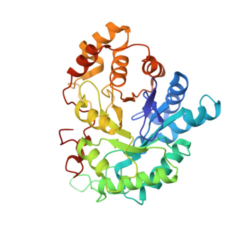
    The crystal structure of a novel sulfonyl-pyridazinone inhibitor in complex with aldose reductase, the first enzyme of the polyol pathway, has been determined to 1.43 angstroms and 0.95 angstroms resolution. The ternary complex of inhibitor, cofactor and enzyme has been obtained by soaking of preformed crystals. Supposedly due to low solubility in the crystallisation buffer, in both structures the inhibitor shows reduced occupancy of 74% and 46% population, respectively. The pyridazinone head group of the inhibitor occupies the catalytic site, whereas the chloro-benzofuran moiety penetrates into the opened specificity pocket. The high-resolution structure provides some evidence that the pyridazinone group binds in a negatively charged deprotonated state, whereas the neighbouring His110 residue most likely adopts a neutral uncharged status. Since the latter structure is populated by the ligand to only 46%, a second conformation of the C-terminal ligand-binding region can be detected. This conformation corresponds to the closed state of the specificity pocket when no or only small ligands are bound to aldose reductase. The two conformational states are in good agreement with frames observed along a molecular dynamics trajectory describing the transition from closed to open situation. Accordingly, both geometries, superimposed in the averaged crystal structure, correspond to snapshots of the ligand-bound and the unbound state. Isothermal titration calorimetry has been applied to determine the binding constants of the investigated pyridazinone in comparison to the hydantoin sorbinil and the carboxylate-type inhibitors IDD 594 and tolrestat. The pyridazinone exhibits a binding affinity similar to those of tolrestat and sorbinil, and shows slightly reduced affinity compared to IDD 594. These studies elucidating the binding mode and providing information about protonation states of protein side-chains involved in binding of this novel class of inhibitors establish the platform for further structure-based drug design.


  • Organizational Affiliation

    Department of Pharmaceutical Chemistry, Philipps-University Marburg, Marbacher Weg 6, 35032 Marburg, Germany.


Macromolecules
Find similar proteins by:  (by identity cutoff)  |  3D Structure
Entity ID: 1
MoleculeChains Sequence LengthOrganismDetailsImage
aldose reductase316Homo sapiensMutation(s): 0 
EC: 1.1.1.21
UniProt & NIH Common Fund Data Resources
Find proteins for P15121 (Homo sapiens)
Explore P15121 
Go to UniProtKB:  P15121
PHAROS:  P15121
GTEx:  ENSG00000085662 
Entity Groups  
Sequence Clusters30% Identity50% Identity70% Identity90% Identity95% Identity100% Identity
UniProt GroupP15121
Sequence Annotations
Expand
  • Reference Sequence
Small Molecules
Ligands 2 Unique
IDChains Name / Formula / InChI Key2D Diagram3D Interactions
NAP
Query on NAP

Download Ideal Coordinates CCD File 
B [auth A]NADP NICOTINAMIDE-ADENINE-DINUCLEOTIDE PHOSPHATE
C21 H28 N7 O17 P3
XJLXINKUBYWONI-NNYOXOHSSA-N
62P
Query on 62P

Download Ideal Coordinates CCD File 
C [auth A]6-[(5-CHLORO-3-METHYL-1-BENZOFURAN-2-YL)SULFONYL]PYRIDAZIN-3(2H)-ONE
C13 H9 Cl N2 O4 S
FXFPQPNUMWQRAO-UHFFFAOYSA-N
Binding Affinity Annotations 
IDSourceBinding Affinity
62P Binding MOAD:  1Z8A Ka: 1.10e+6 (M^-1) from 1 assay(s)
BindingDB:  1Z8A IC50: min: 0.84, max: 840 (nM) from 3 assay(s)
-TΔS: min: -1.25e+1, max: 36.89 (kJ/mol) from 3 assay(s)
ΔH: -5.40e+1 (kJ/mol) from 1 assay(s)
ΔG: min: -4.22e+1, max: -3.45e+1 (kJ/mol) from 4 assay(s)
Experimental Data & Validation

Experimental Data

  • Method: X-RAY DIFFRACTION
  • Resolution: 0.95 Å
  • R-Value Free: 0.142 
  • R-Value Work: 0.122 
  • R-Value Observed: 0.122 
  • Space Group: P 1 21 1
Unit Cell:
Length ( Å )Angle ( ˚ )
a = 49.39α = 90
b = 66.86β = 91.88
c = 47.37γ = 90
Software Package:
Software NamePurpose
SHELXmodel building
SHELXL-97refinement
MAR345data collection
SCALEPACKdata scaling
CNSphasing

Structure Validation

View Full Validation Report


Ligand Structure Quality Assessment 


Entry History 

Deposition Data

Revision History  (Full details and data files)

  • Version 1.0: 2006-03-14
    Type: Initial release
  • Version 1.1: 2008-04-30
    Changes: Version format compliance
  • Version 1.2: 2011-07-13
    Changes: Version format compliance
  • Version 1.3: 2017-10-11
    Changes: Refinement description
  • Version 1.4: 2023-08-23
    Changes: Data collection, Database references, Derived calculations, Refinement description
  • Version 1.5: 2024-03-13
    Changes: Source and taxonomy, Structure summary