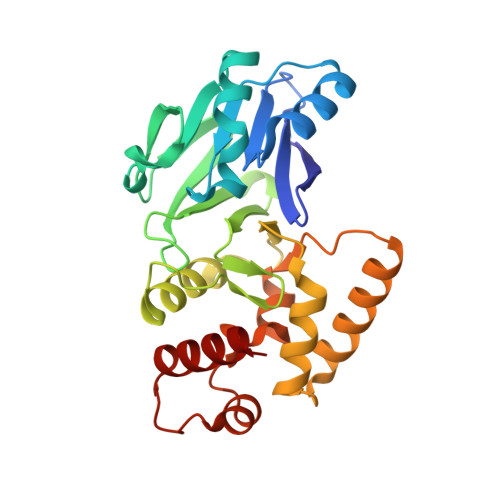
1XM8

X-RAY STRUCTURE OF GLYOXALASE II FROM ARABIDOPSIS THALIANA GENE AT2G31350


Experimental Data Snapshot

  • Method: X-RAY DIFFRACTION
  • Resolution: 1.74 Å
  • R-Value Free: 0.189 
  • R-Value Work: 0.140 
  • R-Value Observed: 0.142 

wwPDB Validation   3D Report Full Report


This is version 1.4 of the entry. See complete history

Re-refinement Note

A newer entry is available that reflects an alternative modeling of the original data: 2Q42


Literature

Structural studies on a mitochondrial glyoxalase II.

Marasinghe, G.P.Sander, I.M.Bennett, B.Periyannan, G.Yang, K.W.Makaroff, C.A.Crowder, M.W.

(2005) J Biol Chem 280: 40668-40675

  • DOI: https://doi.org/10.1074/jbc.M509748200
  • Primary Citation of Related Structures:  
    1XM8

  • PubMed Abstract: 

    Glyoxalase 2 is a beta-lactamase fold-containing enzyme that appears to be involved with cellular chemical detoxification. Although the cytoplasmic isozyme has been characterized from several organisms, essentially nothing is known about the mitochondrial proteins. As a first step in understanding the structure and function of mitochondrial glyoxalase 2 enzymes, a mitochondrial isozyme (GLX2-5) from Arabidopsis thaliana was cloned, overexpressed, purified, and characterized using metal analyses, EPR and (1)H NMR spectroscopies, and x-ray crystallography. The recombinant enzyme was shown to bind 1.04 +/- 0.15 eq of iron and 1.31 +/- 0.05 eq of Zn(II) and to exhibit k(cat) and K(m) values of 129 +/- 10 s(-1) and 391 +/- 48 microm, respectively, when using S-d-lactoylglutathione as the substrate. EPR spectra revealed that recombinant GLX2-5 contains multiple metal centers, including a predominant Fe(III)Z-n(II) center and an anti-ferromagnetically coupled Fe(III)Fe(II) center. Unlike cytosolic glyoxalase 2 from A. thaliana, GLX2-5 does not appear to specifically bind manganese. (1)H NMR spectra revealed the presence of at least eight paramagnetically shifted resonances that arise from protons in close proximity to a Fe(III)Fe(II) center. Five of these resonances arose from solvent-exchangeable protons, and four of these have been assigned to NH protons on metal-bound histidines. A 1.74-A resolution crystal structure of the enzyme revealed that although GLX2-5 shares a number of structural features with human GLX2, several important differences exist. These data demonstrate that mitochondrial glyoxalase 2 can accommodate a number of different metal centers and that the predominant metal center is Fe(III)Zn(II).


  • Organizational Affiliation

    Department of Chemistry and Biochemistry, Miami University, Oxford, Ohio 45056, USA.


Macromolecules
Find similar proteins by:  (by identity cutoff)  |  3D Structure
Entity ID: 1
MoleculeChains Sequence LengthOrganismDetailsImage
glyoxalase II
A, B
254Arabidopsis thalianaMutation(s): 0 
Gene Names: At2g31350
EC: 3.1.2.6
UniProt
Find proteins for Q9SID3 (Arabidopsis thaliana)
Explore Q9SID3 
Go to UniProtKB:  Q9SID3
Entity Groups  
Sequence Clusters30% Identity50% Identity70% Identity90% Identity95% Identity100% Identity
UniProt GroupQ9SID3
Sequence Annotations
Expand
  • Reference Sequence
Small Molecules
Ligands 4 Unique
IDChains Name / Formula / InChI Key2D Diagram3D Interactions
PEG
Query on PEG

Download Ideal Coordinates CCD File 
F [auth A]DI(HYDROXYETHYL)ETHER
C4 H10 O3
MTHSVFCYNBDYFN-UHFFFAOYSA-N
ZN
Query on ZN

Download Ideal Coordinates CCD File 
C [auth A],
G [auth B]
ZINC ION
Zn
PTFCDOFLOPIGGS-UHFFFAOYSA-N
ACY
Query on ACY

Download Ideal Coordinates CCD File 
E [auth A],
I [auth B]
ACETIC ACID
C2 H4 O2
QTBSBXVTEAMEQO-UHFFFAOYSA-N
FE
Query on FE

Download Ideal Coordinates CCD File 
D [auth A],
H [auth B]
FE (III) ION
Fe
VTLYFUHAOXGGBS-UHFFFAOYSA-N
Experimental Data & Validation

Experimental Data

  • Method: X-RAY DIFFRACTION
  • Resolution: 1.74 Å
  • R-Value Free: 0.189 
  • R-Value Work: 0.140 
  • R-Value Observed: 0.142 
  • Space Group: P 1 21 1
Unit Cell:
Length ( Å )Angle ( ˚ )
a = 68.494α = 90
b = 58.782β = 109.22
c = 69.049γ = 90
Software Package:
Software NamePurpose
DENZOdata reduction
SCALEPACKdata scaling
SOLVEphasing
ARP/wARPmodel building
RESOLVEphasing
REFMACrefinement

Structure Validation

View Full Validation Report


Entry History 

Revision History  (Full details and data files)

  • Version 1.0: 2004-10-12
    Type: Initial release
  • Version 1.1: 2008-02-01
    Changes: Version format compliance
  • Version 1.2: 2011-07-13
    Changes: Version format compliance
  • Version 1.3: 2017-10-11
    Changes: Refinement description
  • Version 1.4: 2024-02-14
    Changes: Data collection, Database references, Derived calculations