1XCP

Crystal Structure of the Nitrogenase Fe protein Phe135Trp with MgADP bound


Experimental Data Snapshot

  • Method: X-RAY DIFFRACTION
  • Resolution: 3.20 Å
  • R-Value Free: 0.322 
  • R-Value Work: 0.226 
  • R-Value Observed: 0.229 

wwPDB Validation   3D Report Full Report


Ligand Structure Quality Assessment 


This is version 2.2 of the entry. See complete history


Literature

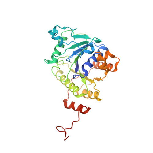
Structural basis for the changes in redox potential in the nitrogenase Phe135Trp Fe protein with MgADP Bound

Jeong, M.S.Jang, S.B.

(2004) Mol Cell 18: 374-382

  • Primary Citation of Related Structures:  
    1XCP

  • PubMed Abstract: 

    The crystal structure of the Azotobacter vinelandii nitrogenase Fe protein with phenylalanine at position 135 substituted by tryptophan has been determined in MgADP-bound form by X-ray diffraction methods. Amino acid substitution studies have suggested that the phenylalanine at position 135 located near the [4Fe-4S] cluster contributes to both the midpoint potential and nucleotide-induced changes of the [4Fe-4S] cluster. Substitution of tryptophan for phenylalanine at position 135 resulted in a significant positive shift in the midpoint potential in both the isolated and nucleotide-bound states. The factors thought to control the midpoint potential of the [FeS] cluster include solvent accessibility, dipolar environment, and structural strain. The structure derived in the present work provides an explanation for the more positive midpoint potential observed in the nucleotide-bound state, and suggests important insights into the contributions of the nucleotide interaction to the conformational states that are the keys to nitrogenase catalysis. The presence of MgADP in Phe135Trp Fe protein reveals the mechanism of the long-range communication from the nucleotide-binding site that controls its affinity for the MoFe protein component.


  • Organizational Affiliation

    Korea Nanobiotechnology Center, Pusan National University, Busan 609-735, Korea.


Macromolecules
Find similar proteins by:  (by identity cutoff)  |  3D Structure
Entity ID: 1
MoleculeChains Sequence LengthOrganismDetailsImage
Nitrogenase iron protein 1
A, B, C, D
289Azotobacter vinelandiiMutation(s): 1 
EC: 1.18.6.1
UniProt
Find proteins for P00459 (Azotobacter vinelandii)
Explore P00459 
Go to UniProtKB:  P00459
Entity Groups  
Sequence Clusters30% Identity50% Identity70% Identity90% Identity95% Identity100% Identity
UniProt GroupP00459
Sequence Annotations
Expand
  • Reference Sequence
Small Molecules
Ligands 3 Unique
IDChains Name / Formula / InChI Key2D Diagram3D Interactions
ADP
Query on ADP

Download Ideal Coordinates CCD File 
F [auth A],
I [auth B],
L [auth C],
N [auth D]
ADENOSINE-5'-DIPHOSPHATE
C10 H15 N5 O10 P2
XTWYTFMLZFPYCI-KQYNXXCUSA-N
SF4
Query on SF4

Download Ideal Coordinates CCD File 
H [auth B],
K [auth C]
IRON/SULFUR CLUSTER
Fe4 S4
LJBDFODJNLIPKO-UHFFFAOYSA-N
MG
Query on MG

Download Ideal Coordinates CCD File 
E [auth A],
G [auth B],
J [auth C],
M [auth D]
MAGNESIUM ION
Mg
JLVVSXFLKOJNIY-UHFFFAOYSA-N
Experimental Data & Validation

Experimental Data

  • Method: X-RAY DIFFRACTION
  • Resolution: 3.20 Å
  • R-Value Free: 0.322 
  • R-Value Work: 0.226 
  • R-Value Observed: 0.229 
  • Space Group: P 21 21 21
Unit Cell:
Length ( Å )Angle ( ˚ )
a = 108.95α = 90
b = 91.26β = 90
c = 125.31γ = 90
Software Package:
Software NamePurpose
DENZOdata reduction
SCALEPACKdata scaling
AMoREphasing
X-PLORrefinement

Structure Validation

View Full Validation Report


Ligand Structure Quality Assessment 


Entry History 

Deposition Data

Revision History  (Full details and data files)

  • Version 1.0: 2005-03-15
    Type: Initial release
  • Version 1.1: 2008-04-30
    Changes: Version format compliance
  • Version 1.2: 2011-07-13
    Changes: Version format compliance
  • Version 2.0: 2019-09-04
    Changes: Atomic model, Data collection, Derived calculations
  • Version 2.1: 2019-11-20
    Changes: Derived calculations
  • Version 2.2: 2021-11-10
    Changes: Database references, Derived calculations