1WMY

Crystal Structure of C-type Lectin CEL-I from Cucumaria echinata


Experimental Data Snapshot

  • Method: X-RAY DIFFRACTION
  • Resolution: 2.00 Å
  • R-Value Free: 0.179 
  • R-Value Work: 0.138 
  • R-Value Observed: 0.140 

wwPDB Validation   3D Report Full Report


This is version 1.3 of the entry. See complete history


Literature

Characteristic Recognition of N-Acetylgalactosamine by an Invertebrate C-type Lectin, CEL-I, Revealed by X-ray Crystallographic Analysis

Sugawara, H.Kusunoki, M.Kurisu, G.Fujimoto, T.Aoyagi, H.Hatakeyama, T.

(2004) J Biol Chem 279: 45219-45225

  • DOI: https://doi.org/10.1074/jbc.M408840200
  • Primary Citation of Related Structures:  
    1WMY, 1WMZ

  • PubMed Abstract: 

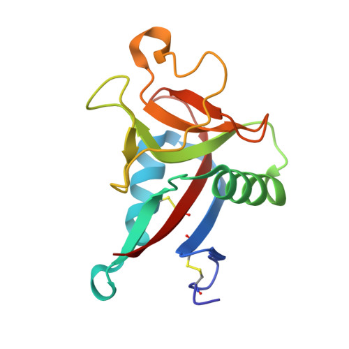
    CEL-I is a C-type lectin, purified from the sea cucumber Cucumaria echinata, that shows a high specificity for N-acetylgalactosamine (GalNAc). We determined the crystal structures of CEL-I and its complex with GalNAc at 2.0 and 1.7 A resolution, respectively. CEL-I forms a disulfide-linked homodimer and contains two intramolecular disulfide bonds, although it lacks one intramolecular disulfide bond that is widely conserved among various C-type carbohydrate recognition domains (CRDs). Although the sequence similarity of CEL-I with other C-type CRDs is low, the overall folding of CEL-I was quite similar to those of other C-type CRDs. The structure of the complex with GalNAc revealed that the basic recognition mode of GalNAc was very similar to that for the GalNAc-binding mutant of the mannose-binding protein. However, the acetamido group of GalNAc appeared to be recognized more strongly by the combination of hydrogen bonds to Arg115 and van der Waals interaction with Gln70. Mutational analyses, in which Gln70 and/or Arg115 were replaced by alanine, confirmed that these residues contributed to GalNAc recognition in a cooperative manner.


  • Organizational Affiliation

    Research Center for Structural and Functional Proteomics, Institute for Protein Research, Osaka University, 3-2 Yamada-oka, Suita, Osaka 565-0871, Japan.


Macromolecules
Find similar proteins by:  (by identity cutoff)  |  3D Structure
Entity ID: 1
MoleculeChains Sequence LengthOrganismDetailsImage
lectin CEL-I, N-acetyl-D-galactosamine-specific C-type
A, B
140Cucumaria echinataMutation(s): 0 
UniProt
Find proteins for Q7M462 (Cucumaria echinata)
Explore Q7M462 
Go to UniProtKB:  Q7M462
Entity Groups  
Sequence Clusters30% Identity50% Identity70% Identity90% Identity95% Identity100% Identity
UniProt GroupQ7M462
Sequence Annotations
Expand
  • Reference Sequence
Experimental Data & Validation

Experimental Data

  • Method: X-RAY DIFFRACTION
  • Resolution: 2.00 Å
  • R-Value Free: 0.179 
  • R-Value Work: 0.138 
  • R-Value Observed: 0.140 
  • Space Group: C 1 2 1
Unit Cell:
Length ( Å )Angle ( ˚ )
a = 92.38α = 90
b = 69.94β = 136.46
c = 76.69γ = 90
Software Package:
Software NamePurpose
REFMACrefinement
DPSdata reduction
CCP4data scaling
AMoREphasing

Structure Validation

View Full Validation Report


Entry History 

Deposition Data

Revision History  (Full details and data files)

  • Version 1.0: 2004-09-07
    Type: Initial release
  • Version 1.1: 2008-04-30
    Changes: Version format compliance
  • Version 1.2: 2011-07-13
    Changes: Advisory, Refinement description, Version format compliance
  • Version 1.3: 2023-10-25
    Changes: Data collection, Database references, Derived calculations, Refinement description