The Structure of the Cytoplasmic Domain of Epsl, an Inner Membrane Component of the Type II Secretion System of Vibrio Cholerae: An Unusual Member of the Actin-Like ATPase Superfamily
Abendroth, J., Bagdasarian, M., Sandkvist, M., Hol, W.G.J.(2004) J Mol Biol 344: 619
- PubMed: 15533433
- DOI: https://doi.org/10.1016/j.jmb.2004.09.062
- Primary Citation of Related Structures:
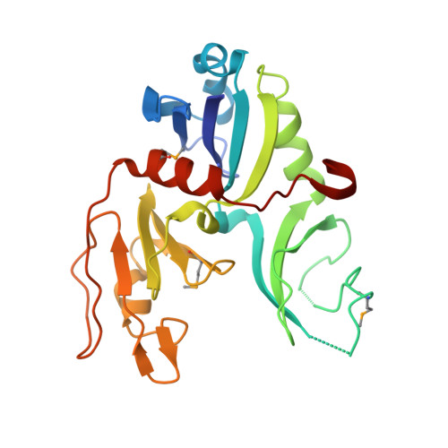
1W97 - PubMed Abstract:
The type II secretion system (T2SS) is used by several Gram-negative bacteria for the secretion of hydrolytic enzymes and virulence factors across the outer membrane. In these secretion systems, a complex of 12-15 so-called "Gsp proteins" spans from a regulatory ATPase in the cytoplasm, via several signal or energy transducing proteins in the inner membrane and the pseudopilins in the periplasm, to the actual pore in the outer membrane. The human pathogen Vibrio cholerae employs such an assembly, called the Eps system, for the export of its major virulence factor, cholera toxin, from its periplasm into the lumen of the gastro-intestinal tract of the host. Here, we report the atomic structure of the major cytoplasmic domain of the inner membrane-spanning EpsL protein from V. cholerae. EpsL is the binding partner of the regulatory ATPase EpsE as well as of EpsM and pseudopilins, and is therefore a critical link between the cytoplasmic and the periplasmic part of the Eps-system. The 2.7A resolution structure was determined by a combination of Se-Met multiple anomalous dispersion (MAD) and multiple isomorphous replacement with anomalous scattering (MIRAS) phasing methods. The 28kDa cytoplasmic domain of EpsL (cyto-EpsL) consists of three beta-sheet-rich domains. With domains I and III similar to the RNaseH-fold, cyto-EpsL unexpectedly shows structural homology with the superfamily of actin-like ATPases. cyto-EpsL, however, is an unusual member of this superfamily as it misses the canonical actin domains 1B and 2B, which are common yet variable in this superfamily. Moreover, cyto-EpsL has an additional domain II, which has the topology of an SHS2-fold module. Within the superfamily this fold module has been observed only for domain 1C of the cell division protein FtsA, in which it mediates protein-protein interactions. This domain II displays great flexibility and contributes to a pronounced negatively charged canyon on the surface of cyto-EpsL. Functional data as well as structural homology and sequence conservation suggest that domain II interacts with EpsE, the major cytoplasmic binding partner of EpsL.
Organizational Affiliation:
Department of Biochemistry, Biomolecular Structure Center, School of Medicine, University of Washington, P.O. Box 357742, Seattle, WA 98195-7242, USA.