1W3U

Crystal structure of phosphoserine aminotransferase from Bacillus circulans var. alkalophilus


Experimental Data Snapshot

  • Method: X-RAY DIFFRACTION
  • Resolution: 1.50 Å
  • R-Value Free: 0.208 
  • R-Value Observed: 0.142 

wwPDB Validation   3D Report Full Report


Ligand Structure Quality Assessment 


This is version 1.4 of the entry. See complete history


Literature

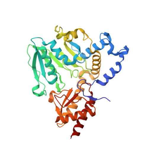
Enzyme Adaptation to Alkaline Ph: Atomic Resolution (1.08 A) Structure of Phosphoserine Aminotransferase from Bacillus Alcalophilus

Dubnovitsky, A.P.Kapetaniou, E.G.Papageorgiou, A.C.

(2005) Protein Sci 14: 97

  • DOI: https://doi.org/10.1110/ps.041029805
  • Primary Citation of Related Structures:  
    1W23, 1W3U

  • PubMed Abstract: 

    The crystal structure of the vitamin B(6)-dependent enzyme phosphoserine aminotransferase from the obligatory alkaliphile Bacillus alcalophilus has been determined at 1.08 A resolution. The model was refined to an R-factor of 11.7% (R(free) = 13.9%). The enzyme displays a narrow pH optimum of enzymatic activity at pH 9.0. The final structure was compared to the previously reported structure of the mesophilic phosphoserine aminotransferase from Escherichia coli and to that of phosphoserine aminotransferase from a facultative alkaliphile, Bacillus circulans subsp. alkalophilus. All three enzymes are homodimers with each monomer comprising a two-domain architecture. Despite the high structural similarity, the alkaliphilic representatives possess a set of distinctive structural features. Two residues directly interacting with pyridoxal-5'-phosphate are replaced, and an additional hydrogen bond to the O3' atom of the cofactor is present in alkaliphilic phosphoserine aminotransferases. The number of hydrogen bonds and hydrophobic interactions at the dimer interface is increased. Hydrophobic interactions between the two domains in the monomers are enhanced. Moreover, the number of negatively charged amino acid residues increases on the solvent-accessible molecular surface and fewer hydrophobic residues are exposed to the solvent. Further, the total amount of ion pairs and ion networks is significantly reduced in the Bacillus enzymes, while the total number of hydrogen bonds is increased. The mesophilic enzyme from Escherichia coli contains two additional beta-strands in a surface loop with a third beta-strand being shorter in the structure. The identified structural features are proposed to be possible factors implicated in the alkaline adaptation of phosphoserine aminotransferase.


  • Organizational Affiliation

    Turku Centre for Biotechnology, University of Turku, Finland.


Macromolecules
Find similar proteins by:  (by identity cutoff)  |  3D Structure
Entity ID: 1
MoleculeChains Sequence LengthOrganismDetailsImage
PHOSPHOSERINE AMINOTRANSFERASE362Niallia circulansMutation(s): 1 
EC: 2.6.1.52
UniProt
Find proteins for Q59196 (Niallia circulans)
Explore Q59196 
Go to UniProtKB:  Q59196
Entity Groups  
Sequence Clusters30% Identity50% Identity70% Identity90% Identity95% Identity100% Identity
UniProt GroupQ59196
Sequence Annotations
Expand
  • Reference Sequence
Small Molecules
Ligands 2 Unique
IDChains Name / Formula / InChI Key2D Diagram3D Interactions
PLP
Query on PLP

Download Ideal Coordinates CCD File 
B [auth A]PYRIDOXAL-5'-PHOSPHATE
C8 H10 N O6 P
NGVDGCNFYWLIFO-UHFFFAOYSA-N
GOL
Query on GOL

Download Ideal Coordinates CCD File 
C [auth A]GLYCEROL
C3 H8 O3
PEDCQBHIVMGVHV-UHFFFAOYSA-N
Experimental Data & Validation

Experimental Data

  • Method: X-RAY DIFFRACTION
  • Resolution: 1.50 Å
  • R-Value Free: 0.208 
  • R-Value Observed: 0.142 
  • Space Group: C 1 2 1
Unit Cell:
Length ( Å )Angle ( ˚ )
a = 91.287α = 90
b = 91.109β = 111.25
c = 42.374γ = 90
Software Package:
Software NamePurpose
SHELXL-97refinement
DENZOdata reduction
SCALEPACKdata scaling
AMoREphasing

Structure Validation

View Full Validation Report


Ligand Structure Quality Assessment 


Entry History 

Deposition Data

Revision History  (Full details and data files)

  • Version 1.0: 2004-12-22
    Type: Initial release
  • Version 1.1: 2013-06-26
    Changes: Database references, Derived calculations, Non-polymer description, Other, Structure summary, Version format compliance
  • Version 1.2: 2019-05-22
    Changes: Data collection, Derived calculations, Other, Refinement description
  • Version 1.3: 2019-07-24
    Changes: Data collection
  • Version 1.4: 2023-12-13
    Changes: Data collection, Database references, Derived calculations, Other, Refinement description