1VCX

Neutron Crystal Structure of the Wild Type Rubredoxin from Pyrococcus Furiosus at 1.5A Resolution


Experimental Data Snapshot

  • Method: NEUTRON DIFFRACTION
  • Resolution: 1.50 Å
  • R-Value Free: 0.217 
  • R-Value Work: 0.186 

wwPDB Validation   3D Report Full Report


This is version 1.6 of the entry. See complete history


Literature

Neutron crystallographic study on rubredoxin from Pyrococcus furiosus by BIX-3, a single-crystal diffractometer for biomacromolecules

Kurihara, K.Tanaka, I.Chatake, T.Adams, M.W.W.Jenney Jr., F.E.Moiseeva, N.Bau, R.Niimura, N.

(2004) Proc Natl Acad Sci U S A 101: 11215-11220

  • DOI: https://doi.org/10.1073/pnas.0403807101
  • Primary Citation of Related Structures:  
    1VCX

  • PubMed Abstract: 

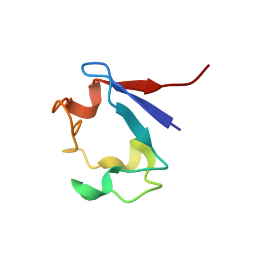
    The structure of a partially deuterated rubredoxin from the hyperthermophilic archaeon Pyrococcus furiosus, an organism that grows optimally at 100 degrees C, was determined by using the neutron single-crystal diffractometer dedicated for biological macromolecules (BIX-3) at the JRR-3M reactor of the Japan Atomic Energy Research Institute. Data were collected at room temperature up to a resolution of 1.5 A, and the completeness factor of the data set was 81.9%. The model contains 306 H and 50 D atoms. A total of 37 hydration water molecules were identified, with 15 having all three atoms fully located and the remaining D2O molecules partially defined. The model has been refined to final agreement factors of R = 18.6% and Rfree = 21.7%. Several orientations of the O-D bonds of side chains, whose assignments from x-ray data were previously ambiguous, were clearly visible in the neutron structure. Although most backbone N-H bonds had undergone some degree of H/D exchange throughout the rubredoxin molecule, 5 H atom positions still had distinctly negative (H) peaks. The neutron Fourier maps clearly showed the details of an extensive set of H bonds involving the ND3+ terminus that may contribute to the unusual thermostability of this molecule.


  • Organizational Affiliation

    Neutron Science Research Center, Japan Atomic Energy Research Institute, Tokai, Ibaraki 319-1195, Japan.


Macromolecules
Find similar proteins by:  (by identity cutoff)  |  3D Structure
Entity ID: 1
MoleculeChains Sequence LengthOrganismDetailsImage
Rubredoxin53Pyrococcus furiosusMutation(s): 0 
UniProt
Find proteins for P24297 (Pyrococcus furiosus (strain ATCC 43587 / DSM 3638 / JCM 8422 / Vc1))
Explore P24297 
Go to UniProtKB:  P24297
Entity Groups  
Sequence Clusters30% Identity50% Identity70% Identity90% Identity95% Identity100% Identity
UniProt GroupP24297
Sequence Annotations
Expand
  • Reference Sequence
Small Molecules
Ligands 1 Unique
IDChains Name / Formula / InChI Key2D Diagram3D Interactions
FE
Query on FE

Download Ideal Coordinates CCD File 
B [auth A]FE (III) ION
Fe
VTLYFUHAOXGGBS-UHFFFAOYSA-N
Experimental Data & Validation

Experimental Data

  • Method: NEUTRON DIFFRACTION
  • Resolution: 1.50 Å
  • R-Value Free: 0.217 
  • R-Value Work: 0.186 
  • Space Group: P 21 21 21
Unit Cell:
Length ( Å )Angle ( ˚ )
a = 34.32α = 90
b = 35.31β = 90
c = 44.23γ = 90
Software Package:
Software NamePurpose
CNSrefinement
DENZOdata reduction
SCALEPACKdata scaling
X-PLORphasing
CNSphasing

Structure Validation

View Full Validation Report


Entry History 

Deposition Data

Revision History  (Full details and data files)

  • Version 1.0: 2004-08-10
    Type: Initial release
  • Version 1.1: 2008-04-27
    Changes: Version format compliance
  • Version 1.2: 2011-07-13
    Changes: Non-polymer description, Version format compliance
  • Version 1.3: 2018-03-07
    Changes: Data collection
  • Version 1.4: 2018-07-11
    Changes: Data collection
  • Version 1.5: 2018-07-18
    Changes: Data collection
  • Version 1.6: 2023-10-25
    Changes: Data collection, Database references, Derived calculations, Refinement description