1VBU

Crystal structure of native xylanase 10B from Thermotoga maritima


Experimental Data Snapshot

  • Method: X-RAY DIFFRACTION
  • Resolution: 1.80 Å
  • R-Value Free: 0.218 
  • R-Value Work: 0.186 
  • R-Value Observed: 0.191 

wwPDB Validation   3D Report Full Report


This is version 1.3 of the entry. See complete history


Literature

Structural basis of the substrate subsite and the highly thermal stability of xylanase 10B from Thermotoga maritima MSB8

IhsanawatiKumasaka, T.Kaneko, T.Morokuma, C.Yatsunami, R.Sato, T.Nakamura, S.Tanaka, N.

(2005) Proteins 61: 999-1009

  • DOI: https://doi.org/10.1002/prot.20700
  • Primary Citation of Related Structures:  
    1VBR, 1VBU

  • PubMed Abstract: 

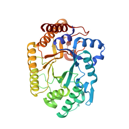
    The crystal structure of xylanase 10B from Thermotoga maritima MSB8 (TmxB), a hyperthermostable xylanase, has been solved in its native form and in complex with xylobiose or xylotriose at 1.8 A resolution. In order to gain insight into the substrate subsite and the molecular features for thermal stability, we compared TmxB with family 10 xylanase structures from nine microorganisms. As expected, TmxB folds into a (beta/alpha)8-barrel structure, which is common among the glycoside hydrolase family 10. The enzyme active site and the environment surrounding the xylooligosaccharide of TmxB are highly similar to those of family 10 xylanases. However, only two xylose moieties were found in its binding pocket from the TmxB-xylotriose complex structure. This finding suggests that TmxB could be a potential biocatalyst for the large-scale production of xylobiose. The result of structural analyses also indicated that TmxB possesses some additional features that account for its thermostability. In particular, clusters of aromatic residues together with a lack of exposed hydrophobic residues are characteristic of the TmxB structure. TmxB has also a significant number of ion pairs on the protein surface that are not found in other thermophilic family 10 xylanases.


  • Organizational Affiliation

    Department of Life Science, Tokyo Institute of Technology, Yokohama, Japan.


Macromolecules
Find similar proteins by:  (by identity cutoff)  |  3D Structure
Entity ID: 1
MoleculeChains Sequence LengthOrganismDetailsImage
endo-1,4-beta-xylanase B
A, B
328Thermotoga maritimaMutation(s): 0 
EC: 3.2.1.8
UniProt
Find proteins for Q9WXS5 (Thermotoga maritima (strain ATCC 43589 / DSM 3109 / JCM 10099 / NBRC 100826 / MSB8))
Explore Q9WXS5 
Go to UniProtKB:  Q9WXS5
Entity Groups  
Sequence Clusters30% Identity50% Identity70% Identity90% Identity95% Identity100% Identity
UniProt GroupQ9WXS5
Sequence Annotations
Expand
  • Reference Sequence
Small Molecules
Ligands 3 Unique
IDChains Name / Formula / InChI Key2D Diagram3D Interactions
SO4
Query on SO4

Download Ideal Coordinates CCD File 
C [auth A]SULFATE ION
O4 S
QAOWNCQODCNURD-UHFFFAOYSA-L
GOL
Query on GOL

Download Ideal Coordinates CCD File 
H [auth A],
K [auth B]
GLYCEROL
C3 H8 O3
PEDCQBHIVMGVHV-UHFFFAOYSA-N
ACY
Query on ACY

Download Ideal Coordinates CCD File 
D [auth A]
E [auth A]
F [auth A]
G [auth A]
I [auth B]
D [auth A],
E [auth A],
F [auth A],
G [auth A],
I [auth B],
J [auth B]
ACETIC ACID
C2 H4 O2
QTBSBXVTEAMEQO-UHFFFAOYSA-N
Experimental Data & Validation

Experimental Data

  • Method: X-RAY DIFFRACTION
  • Resolution: 1.80 Å
  • R-Value Free: 0.218 
  • R-Value Work: 0.186 
  • R-Value Observed: 0.191 
  • Space Group: P 1
Unit Cell:
Length ( Å )Angle ( ˚ )
a = 58.5α = 84.59
b = 59.1β = 71.14
c = 61.34γ = 68.32
Software Package:
Software NamePurpose
CNSrefinement
MOSFLMdata reduction
CCP4data scaling
MOLREPphasing

Structure Validation

View Full Validation Report


Entry History 

Deposition Data

Revision History  (Full details and data files)

  • Version 1.0: 2005-06-28
    Type: Initial release
  • Version 1.1: 2008-04-27
    Changes: Version format compliance
  • Version 1.2: 2011-07-13
    Changes: Non-polymer description, Version format compliance
  • Version 1.3: 2023-12-27
    Changes: Data collection, Database references, Derived calculations