1V29

Crystal structure of Nitrile hydratase from a thermophile Bacillus smithii


Experimental Data Snapshot

  • Method: X-RAY DIFFRACTION
  • Resolution: 2.60 Å
  • R-Value Free: 0.226 
  • R-Value Work: 0.185 
  • R-Value Observed: 0.187 

wwPDB Validation   3D Report Full Report


This is version 1.4 of the entry. See complete history


Literature

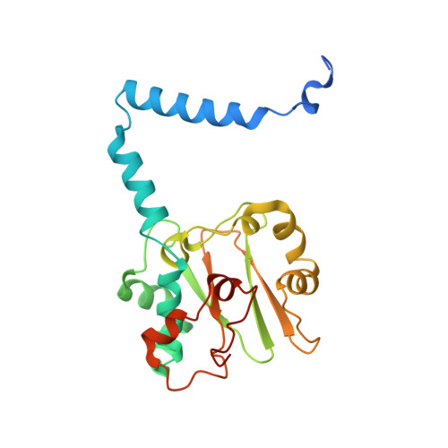
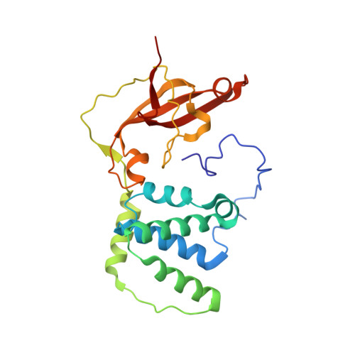
Crystal structure of nitrile hydratase from a thermophilic Bacillus smithii

Hourai, S.Miki, M.Takashima, Y.Mitsuda, S.Yanagi, K.

(2003) Biochem Biophys Res Commun 312: 340-345

  • DOI: https://doi.org/10.1016/j.bbrc.2003.10.124
  • Primary Citation of Related Structures:  
    1V29

  • PubMed Abstract: 

    The crystal structure of the nitrile hydratase (NHase) from Bacillus smithii SC-J05-1 was determined. Our analysis of the structure shows that some residues that seem to be responsible for substrate recognition are different from those of other NHases. In particular, the Phe52 in the beta subunit of NHase from B. smithii covers the metal center partially like a small lid and narrows the active site cleft. It is well known that the NHase from B. smithii especially prefers aliphatic nitriles for its substrate rather than aromatic ones, and we can now infer that the Phe52 residue may play a key role in the substrate specificity for this enzyme. This finding leads us to suggest that substitution of these residues may alter the substrate specificity of the enzyme.


  • Organizational Affiliation

    Sumitomo Chemical Co, Ltd. Environmental Health Science Laboratory, 3-1-98 Kasugade-naka, Konohanaku, Osaka 554-8558, Japan. hourai@sc.sumitomo-chem.co.jp


Macromolecules
Find similar proteins by:  (by identity cutoff)  |  3D Structure
Entity ID: 1
MoleculeChains Sequence LengthOrganismDetailsImage
nitrile hydratase a chain220Bacillus smithiiMutation(s): 0 
EC: 4.2.1.84
UniProt
Find proteins for Q84FS5 (Bacillus sp. RAPc8)
Explore Q84FS5 
Go to UniProtKB:  Q84FS5
Entity Groups  
Sequence Clusters30% Identity50% Identity70% Identity90% Identity95% Identity100% Identity
UniProt GroupQ84FS5
Sequence Annotations
Expand
  • Reference Sequence
Find similar proteins by:  (by identity cutoff)  |  3D Structure
Entity ID: 2
MoleculeChains Sequence LengthOrganismDetailsImage
nitrile hydratase b chain229Bacillus smithiiMutation(s): 0 
EC: 4.2.1.84
Entity Groups  
Sequence Clusters30% Identity50% Identity70% Identity90% Identity95% Identity100% Identity
Sequence Annotations
Expand
  • Reference Sequence
Small Molecules
Ligands 1 Unique
IDChains Name / Formula / InChI Key2D Diagram3D Interactions
CO
Query on CO

Download Ideal Coordinates CCD File 
C [auth A]COBALT (II) ION
Co
XLJKHNWPARRRJB-UHFFFAOYSA-N
Experimental Data & Validation

Experimental Data

  • Method: X-RAY DIFFRACTION
  • Resolution: 2.60 Å
  • R-Value Free: 0.226 
  • R-Value Work: 0.185 
  • R-Value Observed: 0.187 
  • Space Group: I 4 2 2
Unit Cell:
Length ( Å )Angle ( ˚ )
a = 158.42α = 90
b = 158.42β = 90
c = 126.77γ = 90
Software Package:
Software NamePurpose
crystaldata reduction
AMoREphasing
CNXrefinement
CrystalCleardata reduction
CrystalCleardata scaling

Structure Validation

View Full Validation Report


Entry History 

Deposition Data

Revision History  (Full details and data files)

  • Version 1.0: 2004-10-09
    Type: Initial release
  • Version 1.1: 2008-04-27
    Changes: Version format compliance
  • Version 1.2: 2011-07-13
    Changes: Version format compliance
  • Version 1.3: 2017-10-04
    Changes: Refinement description
  • Version 1.4: 2023-12-27
    Changes: Data collection, Database references, Derived calculations