1T3M

Structure of the isoaspartyl peptidase with L-asparaginase activity from E. coli


Experimental Data Snapshot

  • Method: X-RAY DIFFRACTION
  • Resolution: 1.65 Å
  • R-Value Free: 0.217 
  • R-Value Work: 0.174 
  • R-Value Observed: 0.173 

wwPDB Validation   3D Report Full Report


This is version 1.3 of the entry. See complete history


Literature

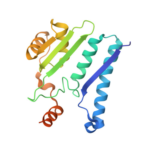
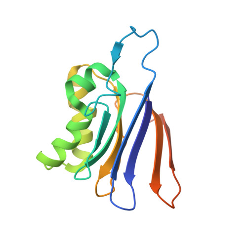
Structure of the isoaspartyl peptidase with L-asparaginase activity from Escherichia coli.

Prahl, A.Pazgier, M.Hejazi, M.Lockau, W.Lubkowski, J.

(2004) Acta Crystallogr D Biol Crystallogr 60: 1173-1176

  • DOI: https://doi.org/10.1107/S0907444904003403
  • Primary Citation of Related Structures:  
    1T3M

  • PubMed Abstract: 

    The crystal structure of the Escherichia coli enzyme (EcAIII) with isoaspartyl dipeptidase and L-asparaginase activity has been solved and refined to a resolution of 1.65 angstroms, with crystallographic R-factor and Rfree values of 0.178 and 0.209, respectively. EcAIII belongs to the family of N-terminal hydrolases. The amino-acid sequence of EcAIII is homologous to those of putative asparaginases from plants. The structure of EcAIII is similar to the structures of glycosylasparaginases. The mature and catalytically active form of EcAIII is a heterotetramer consisting of two alpha-subunits and two beta-subunits. Both of the equivalent active sites present in the EcAIII tetramer is assisted by a metal-binding site. The metal cations, modelled here as Na+, have not previously been observed in glycosylasparaginases. This reported structure helps to explain the inability of EcAIII and other plant-type asparaginases to hydrolyze N4-(beta-N-acetylglucosaminyl)-L-asparagine, the substrate of glycosylasparaginases.


  • Organizational Affiliation

    Macromolecular Crystallography Laboratory, National Cancer Institute at Frederick, Frederick, MD 21702, USA.


Macromolecules
Find similar proteins by:  (by identity cutoff)  |  3D Structure
Entity ID: 1
MoleculeChains Sequence LengthOrganismDetailsImage
Putative L-asparaginase
A, C
177Escherichia coliMutation(s): 0 
Gene Names: YBIKB0828
EC: 3.5.1.1
UniProt
Find proteins for P37595 (Escherichia coli (strain K12))
Explore P37595 
Go to UniProtKB:  P37595
Entity Groups  
Sequence Clusters30% Identity50% Identity70% Identity90% Identity95% Identity100% Identity
UniProt GroupP37595
Sequence Annotations
Expand
  • Reference Sequence
Find similar proteins by:  (by identity cutoff)  |  3D Structure
Entity ID: 2
MoleculeChains Sequence LengthOrganismDetailsImage
Putative L-asparaginase
B, D
147Escherichia coliMutation(s): 0 
Gene Names: YBIKB0828
EC: 3.5.1.1
UniProt
Find proteins for P37595 (Escherichia coli (strain K12))
Explore P37595 
Go to UniProtKB:  P37595
Entity Groups  
Sequence Clusters30% Identity50% Identity70% Identity90% Identity95% Identity100% Identity
UniProt GroupP37595
Sequence Annotations
Expand
  • Reference Sequence
Experimental Data & Validation

Experimental Data

  • Method: X-RAY DIFFRACTION
  • Resolution: 1.65 Å
  • R-Value Free: 0.217 
  • R-Value Work: 0.174 
  • R-Value Observed: 0.173 
  • Space Group: P 21 21 21
Unit Cell:
Length ( Å )Angle ( ˚ )
a = 66.318α = 90
b = 71.637β = 90
c = 149.583γ = 90
Software Package:
Software NamePurpose
HKL-2000data collection
HKL-2000data reduction
AMoREphasing
SHELXL-97refinement
HKL-2000data scaling

Structure Validation

View Full Validation Report


Entry History 

Deposition Data

Revision History  (Full details and data files)

  • Version 1.0: 2004-07-13
    Type: Initial release
  • Version 1.1: 2008-04-30
    Changes: Version format compliance
  • Version 1.2: 2011-07-13
    Changes: Version format compliance
  • Version 1.3: 2023-08-23
    Changes: Data collection, Database references, Derived calculations, Refinement description