Structure of sesbania mosaic virus at 3 A resolution.
Bhuvaneshwari, M., Subramanya, H.S., Gopinath, K., Savithri, H.S., Nayudu, M.V., Murthy, M.R.(1995) Structure 3: 1021-1030
- PubMed: 8589997
- DOI: https://doi.org/10.1016/s0969-2126(01)00238-6
- Primary Citation of Related Structures:
1SMV - PubMed Abstract:
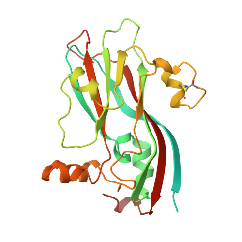
Sobemoviruses are a group of RNA plant viruses that have a narrow host range. They are characterized in vitro by their stability, high thermal inactivation point and longevity. The three-dimensional structure of only one virus belonging to this group, southern bean mosaic virus (SBMV), is known. Structural studies on sesbania mosaic virus (SMV), which is closely related to SBMV, will provide details of the molecular interactions that are likely to be important in the stability and assembly of sobemoviruses. We have determined the three-dimensional structure of SMV at 3 A resolution. The polypeptide fold and quaternary organization are very similar to those of SBMV. The capsid consists of sixty icosahedral asymmetric units, each comprising three copies of a chemically identical coat protein subunit, which are designated as A, B and C and are in structurally different environments. Four cation-binding sites have been located in the icosahedral asymmetric unit. Of these, the site at the quasi-threefold axis is not found in SBMV. Structural differences are observed in loops and regions close to this cation-binding site. Preliminary studies on ethylene diamine tetra acetic acid (EDTA) treated crystals suggest asymmetry in removal of the quasi-equivalent cations at the AB, BC, and AC subunit interfaces. Despite the overall similarity between SMV and SBMV in the nature of the polypeptide fold, these viruses show a number of differences in intermolecular interactions. The polar interactions at the quasi-threefold axis are substantially less in SMV and positively charged residues on the RNA-facing side of the protein and in the N-terminal arm are not particularly well conserved. This suggests that protein-RNA interactions are likely to be different between the two viruses.
Organizational Affiliation:
Molecular Biophysics Unit, Indian Institute of Science, Bangalore, India.