1SGZ

Crystal Structure of Unbound Beta-Secretase Catalytic Domain.


Experimental Data Snapshot

  • Method: X-RAY DIFFRACTION
  • Resolution: 2.00 Å
  • R-Value Free: 0.228 
  • R-Value Work: 0.196 
  • R-Value Observed: 0.196 

wwPDB Validation   3D Report Full Report


This is version 1.3 of the entry. See complete history


Literature

Flap Position of Free Memapsin 2 (beta-Secretase), a Model for Flap Opening in Aspartic Protease Catalysis

Hong, L.Tang, J.

(2004) Biochemistry 43: 4689-4695

  • DOI: https://doi.org/10.1021/bi0498252
  • Primary Citation of Related Structures:  
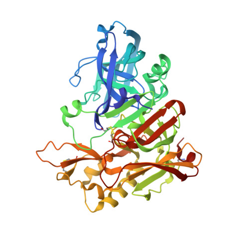
    1SGZ

  • PubMed Abstract: 

    The three-dimensional structure of unbound human memapsin 2 (beta-secretase) protease domain determined at 2.0-A resolution has revealed a new position of the flap region, which appears to be locked in an "open" position. While the structure outside of the flap is essentially the same as the structure of memapsin 2 bound to an inhibitor, the flap positions are 4.5 A different at the tips. The open position of the flap in the current structure is stabilized by two newly formed intraflap hydrogen bonds and anchored by a new hydrogen bond involving the side chain of Tyr 71 (Tyr 75 in pepsin numbering) in a novel orientation. In molecular modeling experiments, the opening of the flap, 6.5 A at the narrowest point, permits entrance of substrates into the cleft. The narrowest point of the opening may function to discriminate among substrates based on sequence and shape. The observed flap opening may also serve as a model for the flap movement in the catalytic mechanism of eukaryotic aspartic proteases and provide insight for the side-chain selection in the design of memapsin 2 inhibitors.


  • Organizational Affiliation

    Protein Studies Program, Oklahoma Medical Research Foundation, Oklahoma City, Oklahoma 73104, USA.


Macromolecules
Find similar proteins by:  (by identity cutoff)  |  3D Structure
Entity ID: 1
MoleculeChains Sequence LengthOrganismDetailsImage
Beta-secretase
A, B, C, D
389Homo sapiensMutation(s): 0 
Gene Names: BACEBACE1
EC: 3.4.23.46
UniProt & NIH Common Fund Data Resources
Find proteins for P56817 (Homo sapiens)
Explore P56817 
Go to UniProtKB:  P56817
PHAROS:  P56817
GTEx:  ENSG00000186318 
Entity Groups  
Sequence Clusters30% Identity50% Identity70% Identity90% Identity95% Identity100% Identity
UniProt GroupP56817
Sequence Annotations
Expand
  • Reference Sequence
Experimental Data & Validation

Experimental Data

  • Method: X-RAY DIFFRACTION
  • Resolution: 2.00 Å
  • R-Value Free: 0.228 
  • R-Value Work: 0.196 
  • R-Value Observed: 0.196 
  • Space Group: P 1 21 1
Unit Cell:
Length ( Å )Angle ( ˚ )
a = 86.16α = 90
b = 130.812β = 97.27
c = 88.651γ = 90
Software Package:
Software NamePurpose
DENZOdata reduction
SCALEPACKdata scaling
AMoREphasing
CNSrefinement

Structure Validation

View Full Validation Report


Entry History 

Deposition Data

Revision History  (Full details and data files)

  • Version 1.0: 2004-05-04
    Type: Initial release
  • Version 1.1: 2008-04-29
    Changes: Version format compliance
  • Version 1.2: 2011-07-13
    Changes: Version format compliance
  • Version 1.3: 2023-08-23
    Changes: Data collection, Database references, Refinement description