Crystal structures of HIV protease V82A and L90M mutants reveal changes in the indinavir-binding site
Mahalingam, B., Wang, Y.-F., Boross, P.I., Tozser, J., Louis, J.M., Harrison, R.W., Weber, I.T.(2004) Eur J Biochem 271: 1516-1524
- PubMed: 15066177
- DOI: https://doi.org/10.1111/j.1432-1033.2004.04060.x
- Primary Citation of Related Structures:
1SDT, 1SDU, 1SDV - PubMed Abstract:
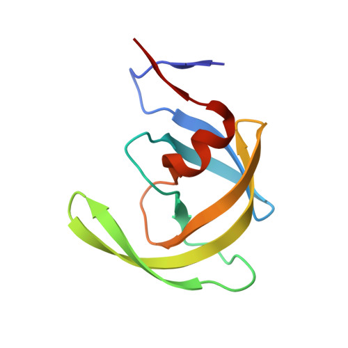
The crystal structures of the wild-type HIV-1 protease (PR) and the two resistant variants, PR(V82A) and PR(L90M), have been determined in complex with the antiviral drug, indinavir, to gain insight into the molecular basis of drug resistance. V82A and L90M correspond to an active site mutation and nonactive site mutation, respectively. The inhibition (K(i)) of PR(V82A) and PR(L90M) was 3.3- and 0.16-fold, respectively, relative to the value for PR. They showed only a modest decrease, of 10-15%, in their k(cat)/K(m) values relative to PR. The crystal structures were refined to resolutions of 1.25-1.4 A to reveal critical features associated with inhibitor resistance. PR(V82A) showed local changes in residues 81-82 at the site of the mutation, while PR(L90M) showed local changes near Met90 and an additional interaction with indinavir. These structural differences concur with the kinetic data.
Organizational Affiliation:
Department of Biology, Georgia State University, PO Box 4010, Atlanta, GA 30302-4010, USA.