1N4U

CHOLESTEROL OXIDASE FROM STREPTOMYCES @ pH 4.5 (STREPTOMYCES SP. SA-COO)


Experimental Data Snapshot

  • Method: X-RAY DIFFRACTION
  • Resolution: 0.95 Å
  • R-Value Free: 0.136 
  • R-Value Work: 0.113 

wwPDB Validation   3D Report Full Report


This is version 1.3 of the entry. See complete history


Literature

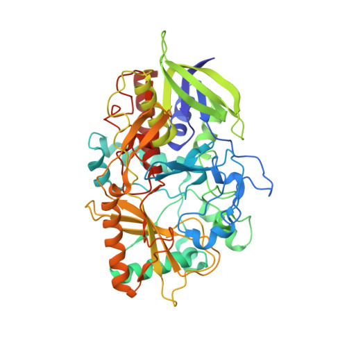
Atomic resolution crystallography reveals how changes in pH shape the protein microenvironment

Lyubimov, A.Y.Lario, P.I.Moustafa, I.Vrielink, A.

(2006) Nat Chem Biol 2: 259-264

  • DOI: https://doi.org/10.1038/nchembio784
  • Primary Citation of Related Structures:  
    1N4U, 1N4V, 1N4W, 2GEW

  • PubMed Abstract: 

    Hydrogen atoms are a vital component of enzyme structure and function. In recent years, atomic resolution crystallography (>or=1.2 A) has been successfully used to investigate the role of the hydrogen atom in enzymatic catalysis. Here, atomic resolution crystallography was used to study the effect of pH on cholesterol oxidase from Streptomyces sp., a flavoenzyme oxidoreductase. Crystallographic observations of the anionic oxidized flavin cofactor at basic pH are consistent with the UV-visible absorption profile of the enzyme and readily explain the reversible pH-dependent loss of oxidation activity. Furthermore, a hydrogen atom, positioned at an unusually short distance from the main chain carbonyl oxygen of Met122 at high pH, was observed, suggesting a previously unknown mechanism of cofactor stabilization. This study shows how a redox active site responds to changes in the enzyme's environment and how these changes are able to influence the mechanism of enzymatic catalysis.


  • Organizational Affiliation

    Department of Molecular, Cell and Developmental Biology, 1156 High Street, University of California, Santa Cruz, California 95064, USA.


Macromolecules
Find similar proteins by:  (by identity cutoff)  |  3D Structure
Entity ID: 1
MoleculeChains Sequence LengthOrganismDetailsImage
Cholesterol oxidase504Streptomyces sp. SA-COOMutation(s): 0 
Gene Names: CHOA
EC: 1.1.3.6
UniProt
Find proteins for P12676 (Streptomyces sp. (strain SA-COO))
Explore P12676 
Go to UniProtKB:  P12676
Entity Groups  
Sequence Clusters30% Identity50% Identity70% Identity90% Identity95% Identity100% Identity
UniProt GroupP12676
Sequence Annotations
Expand
  • Reference Sequence
Experimental Data & Validation

Experimental Data

  • Method: X-RAY DIFFRACTION
  • Resolution: 0.95 Å
  • R-Value Free: 0.136 
  • R-Value Work: 0.113 
  • Space Group: P 1 21 1
Unit Cell:
Length ( Å )Angle ( ˚ )
a = 51.248α = 90
b = 73.276β = 105.07
c = 63.098γ = 90
Software Package:
Software NamePurpose
DENZOdata reduction
SCALEPACKdata scaling
SHELXmodel building
SHELXL-97refinement
SHELXphasing

Structure Validation

View Full Validation Report


Entry History 

Deposition Data

Revision History  (Full details and data files)

  • Version 1.0: 2004-04-27
    Type: Initial release
  • Version 1.1: 2008-04-28
    Changes: Version format compliance
  • Version 1.2: 2011-07-13
    Changes: Source and taxonomy, Version format compliance
  • Version 1.3: 2024-02-14
    Changes: Data collection, Database references, Derived calculations, Refinement description