1MHQ

Crystal Structure Of Human GGA2 VHS Domain


Experimental Data Snapshot

  • Method: X-RAY DIFFRACTION
  • Resolution: 2.20 Å
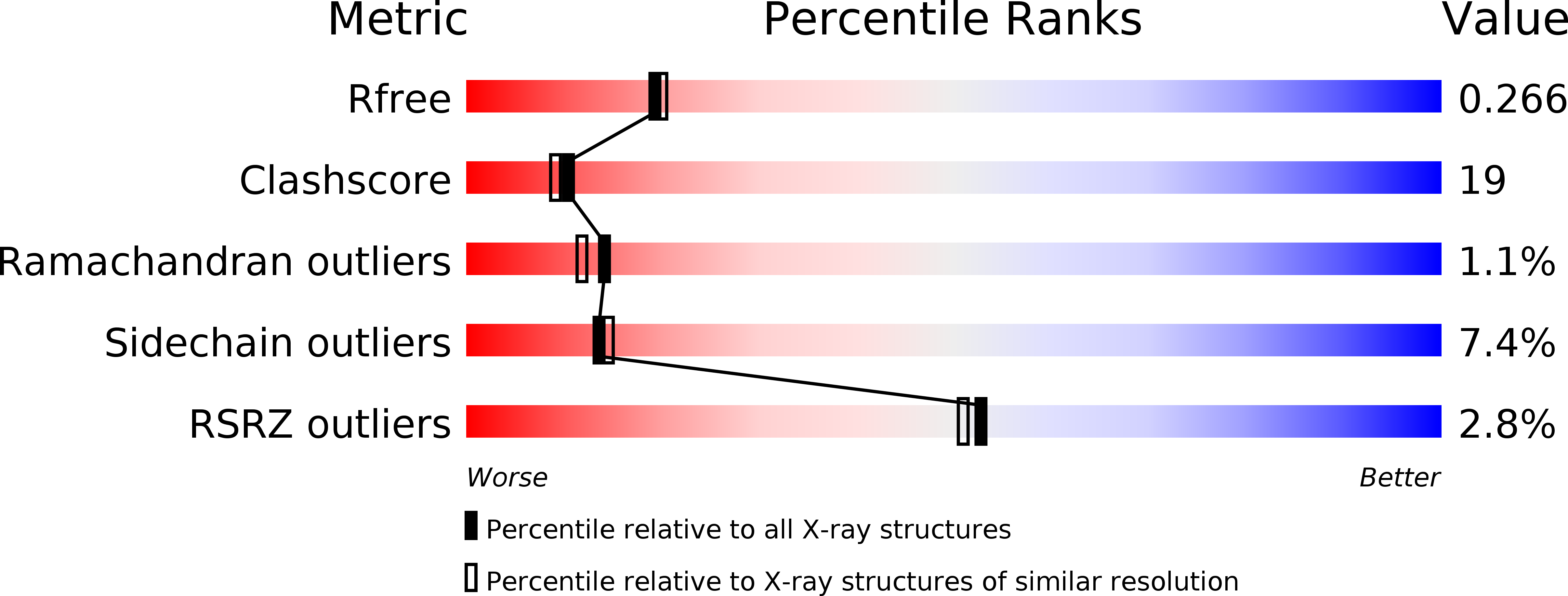
  • R-Value Free: 0.267 
  • R-Value Work: 0.223 
  • R-Value Observed: 0.224 

wwPDB Validation   3D Report Full Report


This is version 1.3 of the entry. See complete history


Literature

Crystal structure of GGA2 VHS domain and its implication in plasticity in the ligand binding pocket

Zhu, G.He, X.Terzyan, S.Zhai, P.Tang, J.Zhang, X.C.

(2003) FEBS Lett 537: 171-176

  • DOI: https://doi.org/10.1016/s0014-5793(03)00095-4
  • Primary Citation of Related Structures:  
    1MHQ

  • PubMed Abstract: 

    Golgi-localized, gamma-ear-containing, ARF binding (GGA) proteins regulate intracellular vesicle transport by recognizing sorting signals on the cargo surface in the initial step of the budding process. The VHS (VPS27, Hrs, and STAM) domain of GGA binds with the signal peptides. Here, a crystal structure of the VHS domain of GGA2 is reported at 2.2 A resolution, which permits a direct comparison with that of homologous proteins, GGA1 and GGA3. Significant structural difference is present in the loop between helices 6 and 7, which forms part of the ligand binding pocket. Intrinsic fluorescence spectroscopic study indicates that this loop undergoes a conformational change upon ligand binding. Thus, the current structure suggests that a conformational change induced by ligand binding occurs in this part of the ligand pocket.


  • Organizational Affiliation

    Crystallography Research Program of Oklahoma Medical Research Foundation, 825 N.E. 13th Street, Oklahoma City 73104, USA.


Macromolecules
Find similar proteins by:  (by identity cutoff)  |  3D Structure
Entity ID: 1
MoleculeChains Sequence LengthOrganismDetailsImage
ADP-ribosylation factor binding protein GGA2
A, B
148Homo sapiensMutation(s): 0 
Membrane Entity: Yes 
UniProt & NIH Common Fund Data Resources
Find proteins for Q9UJY4 (Homo sapiens)
Explore Q9UJY4 
Go to UniProtKB:  Q9UJY4
PHAROS:  Q9UJY4
GTEx:  ENSG00000103365 
Entity Groups  
Sequence Clusters30% Identity50% Identity70% Identity90% Identity95% Identity100% Identity
UniProt GroupQ9UJY4
Sequence Annotations
Expand
  • Reference Sequence
Experimental Data & Validation

Experimental Data

  • Method: X-RAY DIFFRACTION
  • Resolution: 2.20 Å
  • R-Value Free: 0.267 
  • R-Value Work: 0.223 
  • R-Value Observed: 0.224 
  • Space Group: P 21 21 21
Unit Cell:
Length ( Å )Angle ( ˚ )
a = 62.86α = 90
b = 68.11β = 90
c = 74.2γ = 90
Software Package:
Software NamePurpose
MAR345data collection
SCALEPACKdata scaling
AMoREphasing
CNSrefinement

Structure Validation

View Full Validation Report


Entry History 

Deposition Data

Revision History  (Full details and data files)

  • Version 1.0: 2003-03-11
    Type: Initial release
  • Version 1.1: 2008-04-28
    Changes: Version format compliance
  • Version 1.2: 2011-07-13
    Changes: Version format compliance
  • Version 1.3: 2017-10-11
    Changes: Advisory, Refinement description