1MAA

MOUSE ACETYLCHOLINESTERASE CATALYTIC DOMAIN, GLYCOSYLATED PROTEIN


Experimental Data Snapshot

  • Method: X-RAY DIFFRACTION
  • Resolution: 2.90 Å
  • R-Value Free: 0.248 
  • R-Value Work: 0.200 
  • R-Value Observed: 0.200 

wwPDB Validation   3D Report Full Report


Ligand Structure Quality Assessment 


This is version 2.2 of the entry. See complete history


Literature

Crystal structure of mouse acetylcholinesterase. A peripheral site-occluding loop in a tetrameric assembly.

Bourne, Y.Taylor, P.Bougis, P.E.Marchot, P.

(1999) J Biol Chem 274: 2963-2970

  • DOI: https://doi.org/10.1074/jbc.274.5.2963
  • Primary Citation of Related Structures:  
    1MAA

  • PubMed Abstract: 

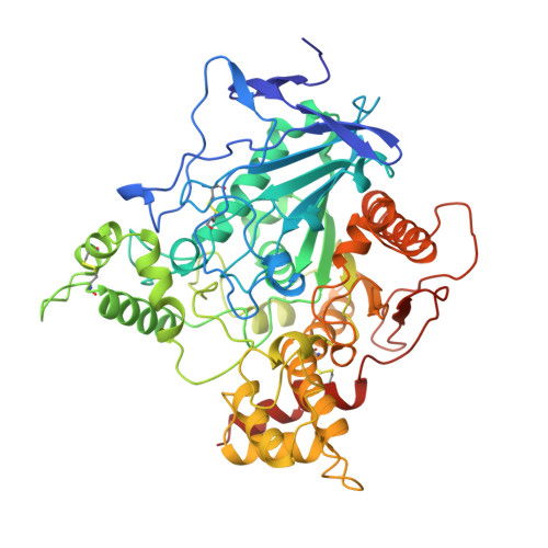
    The crystal structure of mouse acetylcholinesterase at 2.9-A resolution reveals a tetrameric assembly of subunits with an antiparallel alignment of two canonical homodimers assembled through four-helix bundles. In the tetramer, a short Omega loop, composed of a cluster of hydrophobic residues conserved in mammalian acetylcholinesterases along with flanking alpha-helices, associates with the peripheral anionic site of the facing subunit and sterically occludes the entrance of the gorge leading to the active center. The inverse loop-peripheral site interaction occurs within the second pair of subunits, but the peripheral sites on the two loop-donor subunits remain freely accessible to the solvent. The position and complementarity of the peripheral site-occluding loop mimic the characteristics of the central loop of the peptidic inhibitor fasciculin bound to mouse acetylcholinesterase. Tetrameric forms of cholinesterases are widely distributed in nature and predominate in mammalian brain. This structure reveals a likely mode of subunit arrangement and suggests that the peripheral site, located near the rim of the gorge, is a site for association of neighboring subunits or heterologous proteins with interactive surface loops.


  • Organizational Affiliation

    CNRS, Unité Propre de Recherche 9039, Architecture et Fonction des Macromolécules Biologiques, Institut de Biologie et Microbiologie Structurale, F-13402 Marseille Cedex 20, France.


Macromolecules
Find similar proteins by:  (by identity cutoff)  |  3D Structure
Entity ID: 1
MoleculeChains Sequence LengthOrganismDetailsImage
ACETYLCHOLINESTERASE
A, B, C, D
547Mus musculusMutation(s): 0 
Gene Names: MOUSE ACHE
EC: 3.1.1.7
UniProt & NIH Common Fund Data Resources
Find proteins for P21836 (Mus musculus)
Explore P21836 
Go to UniProtKB:  P21836
IMPC:  MGI:87876
Entity Groups  
Sequence Clusters30% Identity50% Identity70% Identity90% Identity95% Identity100% Identity
UniProt GroupP21836
Sequence Annotations
Expand
  • Reference Sequence
Oligosaccharides

Help

Entity ID: 2
MoleculeChains Length2D Diagram Glycosylation3D Interactions
2-acetamido-2-deoxy-beta-D-glucopyranose-(1-4)-[beta-L-fucopyranose-(1-6)]2-acetamido-2-deoxy-beta-D-glucopyranose
E
3N-Glycosylation
Glycosylation Resources
GlyTouCan:  G28454KX
GlyCosmos:  G28454KX
GlyGen:  G28454KX
Small Molecules
Ligands 4 Unique
IDChains Name / Formula / InChI Key2D Diagram3D Interactions
DME
Query on DME

Download Ideal Coordinates CCD File 
G [auth A],
J [auth B],
M [auth C],
P [auth D]
DECAMETHONIUM ION
C16 H38 N2
MTCUAOILFDZKCO-UHFFFAOYSA-N
NAG
Query on NAG

Download Ideal Coordinates CCD File 
F [auth A]2-acetamido-2-deoxy-beta-D-glucopyranose
C8 H15 N O6
OVRNDRQMDRJTHS-FMDGEEDCSA-N
PO4
Query on PO4

Download Ideal Coordinates CCD File 
I [auth B],
L [auth C],
N [auth C]
PHOSPHATE ION
O4 P
NBIIXXVUZAFLBC-UHFFFAOYSA-K
GOL
Query on GOL

Download Ideal Coordinates CCD File 
H [auth A],
K [auth B],
O [auth C],
Q [auth D]
GLYCEROL
C3 H8 O3
PEDCQBHIVMGVHV-UHFFFAOYSA-N
Binding Affinity Annotations 
IDSourceBinding Affinity
DME BindingDB:  1MAA Ki: min: 460, max: 3500 (nM) from 2 assay(s)
IC50: min: 1000, max: 5900 (nM) from 2 assay(s)
Experimental Data & Validation

Experimental Data

  • Method: X-RAY DIFFRACTION
  • Resolution: 2.90 Å
  • R-Value Free: 0.248 
  • R-Value Work: 0.200 
  • R-Value Observed: 0.200 
  • Space Group: P 21 21 21
Unit Cell:
Length ( Å )Angle ( ˚ )
a = 136.55α = 90
b = 173.13β = 90
c = 224.25γ = 90
Software Package:
Software NamePurpose
DENZOdata reduction
SCALAdata scaling
X-PLORmodel building
X-PLORrefinement
CCP4data scaling
X-PLORphasing

Structure Validation

View Full Validation Report


Ligand Structure Quality Assessment 


Entry History 

Deposition Data

Revision History  (Full details and data files)

  • Version 1.0: 1999-04-20
    Type: Initial release
  • Version 1.1: 2008-03-24
    Changes: Version format compliance
  • Version 1.2: 2011-07-13
    Changes: Non-polymer description, Version format compliance
  • Version 2.0: 2020-07-29
    Type: Remediation
    Reason: Carbohydrate remediation
    Changes: Atomic model, Data collection, Database references, Derived calculations, Structure summary
  • Version 2.1: 2021-06-02
    Changes: Derived calculations, Structure summary
  • Version 2.2: 2023-08-09
    Changes: Database references, Refinement description