1LXM

Crystal Structure of Streptococcus agalactiae Hyaluronate Lyase Complexed with Hexasaccharide Unit of Hyaluronan


Experimental Data Snapshot

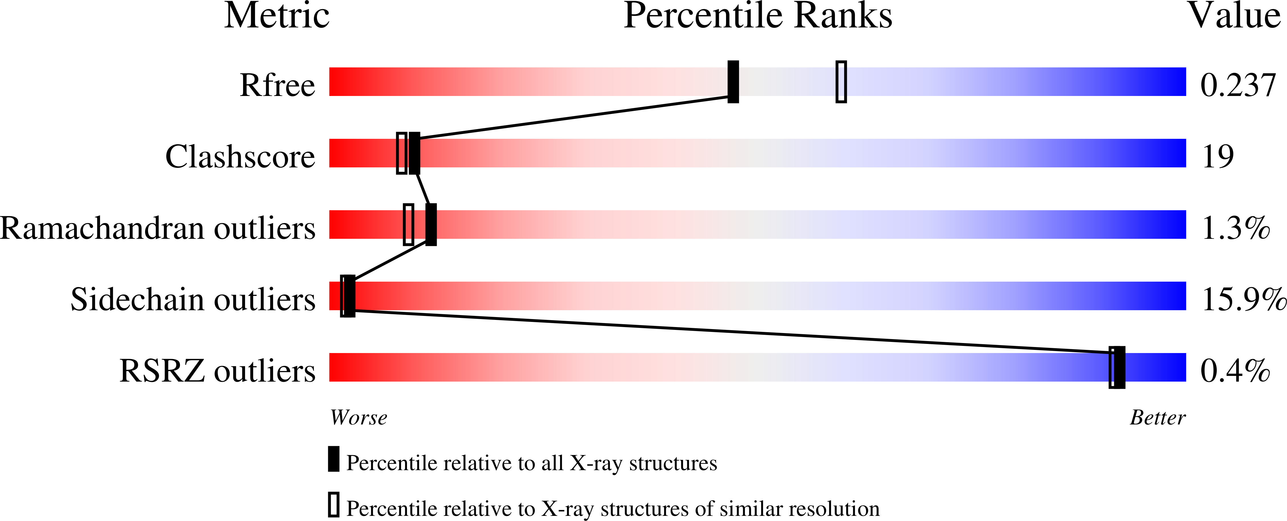
  • Method: X-RAY DIFFRACTION
  • Resolution: 2.20 Å
  • R-Value Free: 0.271 
  • R-Value Work: 0.218 
  • R-Value Observed: 0.218 

wwPDB Validation   3D Report Full Report


This is version 2.1 of the entry. See complete history


Literature

Structure and flexibility of Streptococcus agalactiae hyaluronate lyase complex with its substrate. Insights into the mechanism of processive degradation of hyaluronan.

Mello, L.V.De Groot, B.L.Li, S.Jedrzejas, M.J.

(2002) J Biol Chem 277: 36678-36688

  • DOI: https://doi.org/10.1074/jbc.M205140200
  • Primary Citation of Related Structures:  
    1LXM

  • PubMed Abstract: 

    Streptococcus agalactiae hyaluronate lyase degrades primarily hyaluronan, the main polysaccharide component of the host connective tissues, into unsaturated disaccharide units as the end product. Such function of the enzyme destroys the normal connective tissue structure of the host and exposes the tissue cells to various bacterial toxins. The crystal structure of hexasaccharide hyaluronan complex with the S. agalactiae hyaluronate lyase was determined at 2.2 A resolution; the mechanism of the catalytic process, including the identification of specific residues involved in the degradation of hyaluronan, was clearly identified. The enzyme is composed structurally and functionally from two distinct domains, an alpha-helical alpha-domain and a beta-sheet beta-domain. The flexibility of the protein was investigated by comparing the crystal structures of the S. agalactiae and the Streptococcus pneumoniae enzymes, and by using essential dynamics analyses of CONCOORD computer simulations. These revealed important modes of flexibility, which could be related to the protein function. First, a rotation/twist of the alpha-domain relative to the beta-domain is potentially related to the mechanism of processivity of the enzyme; this twist motion likely facilitates shifting of the ligand along the catalytic site cleft in order to reposition it to be ready for further cleavage. Second, a movement of the alpha- and beta-domains with respect to each other was found to contribute to a change in electrostatic characteristics of the enzyme and appears to facilitate binding of the negatively charged hyaluronan ligand. Third, an opening/closing of the substrate binding cleft brings a catalytic histidine closer to the cleavable substrate beta1,4-glycosidic bond. This opening/closing mode also reflects the main conformational difference between the crystal structures of the S. agalactiae and the S. pneumoniae hyaluronate lyases.


  • Organizational Affiliation

    Children's Hospital Oakland Research Institute, Oakland, California 94609, USA.


Macromolecules
Find similar proteins by:  (by identity cutoff)  |  3D Structure
Entity ID: 1
MoleculeChains Sequence LengthOrganismDetailsImage
HYALURONATE Lyase814Streptococcus agalactiaeMutation(s): 0 
Gene Names: hyl
EC: 4.2.2.1
UniProt
Find proteins for Q53591 (Streptococcus agalactiae serotype III (strain NEM316))
Explore Q53591 
Go to UniProtKB:  Q53591
Entity Groups  
Sequence Clusters30% Identity50% Identity70% Identity90% Identity95% Identity100% Identity
UniProt GroupQ53591
Sequence Annotations
Expand
  • Reference Sequence
Oligosaccharides

Help

Entity ID: 2
MoleculeChains Length2D Diagram Glycosylation3D Interactions
beta-D-glucopyranuronic acid-(1-3)-2-acetamido-2-deoxy-beta-D-glucopyranose-(1-4)-beta-D-glucopyranuronic acid-(1-3)-2-acetamido-2-deoxy-beta-D-glucopyranose-(1-4)-beta-D-glucopyranuronic acid-(1-3)-2-acetamido-2-deoxy-beta-D-glucopyranose
B
6N/A
Glycosylation Resources
GlyTouCan:  G01866TT
GlyCosmos:  G01866TT
GlyGen:  G01866TT
Experimental Data & Validation

Experimental Data

  • Method: X-RAY DIFFRACTION
  • Resolution: 2.20 Å
  • R-Value Free: 0.271 
  • R-Value Work: 0.218 
  • R-Value Observed: 0.218 
  • Space Group: C 2 2 21
Unit Cell:
Length ( Å )Angle ( ˚ )
a = 51.108α = 90
b = 155.043β = 90
c = 238.136γ = 90
Software Package:
Software NamePurpose
HKL-2000data collection
SCALEPACKdata scaling
X-PLORmodel building
X-PLORrefinement
HKL-2000data reduction
X-PLORphasing

Structure Validation

View Full Validation Report


Entry History 

Deposition Data

Revision History  (Full details and data files)

  • Version 1.0: 2002-10-30
    Type: Initial release
  • Version 1.1: 2008-04-28
    Changes: Version format compliance
  • Version 1.2: 2011-07-13
    Changes: Non-polymer description, Version format compliance
  • Version 2.0: 2020-07-29
    Type: Remediation
    Reason: Carbohydrate remediation
    Changes: Advisory, Atomic model, Data collection, Derived calculations, Structure summary
  • Version 2.1: 2024-02-14
    Changes: Advisory, Data collection, Database references, Refinement description, Structure summary