Caught in the act: the structure of phosphorylated beta-phosphoglucomutase from Lactococcus lactis.
Lahiri, S.D., Zhang, G., Dunaway-Mariano, D., Allen, K.N.(2002) Biochemistry 41: 8351-8359
- PubMed: 12081483
- DOI: https://doi.org/10.1021/bi0202373
- Primary Citation of Related Structures:
1LVH - PubMed Abstract:
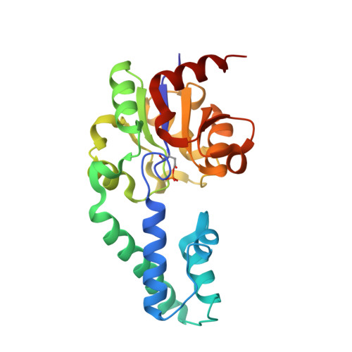
Phosphoglucomutases catalyze the interconversion of D-glucose 1-phosphate and D-glucose 6-phosphate, a reaction central to energy metabolism in all cells and to the synthesis of cell wall polysaccharides in bacterial cells. Two classes of phosphoglucomutases (alpha-PGM and beta-PGM) are distinguished on the basis of their specificity for alpha- and beta-glucose-1-phosphate. beta-PGM is a member of the haloacid dehalogenase (HAD) superfamily, which includes the sarcoplasmic Ca(2+)-ATPase, phosphomannomutase, and phosphoserine phosphatase. beta-PGM is unusual among family members in that the common phosphoenzyme intermediate exists as a stable ground-state complex in this enzyme. Herein we report, for the first time, the three-dimensional structure of a beta-PGM and the first view of the true phosphoenzyme intermediate in the HAD superfamily. The crystal structure of the Mg(II) complex of phosphorylated beta-phosphoglucomutase (beta-PGM) from Lactococcus lactis has been determined to 2.3 A resolution by multiwavelength anomalous diffraction (MAD) phasing on selenomethionine, and refined to an R(cryst) = 0.24 and R(free) = 0.28. The active site of beta-PGM is located between the core and the cap domain and is freely solvent accessible. The residues within a 6 A radius of the phosphorylated Asp8 include Asp10, Thr16, Ser114, Lys145, Glu169, and Asp170. The cofactor Mg(2+) is liganded with octahedral coordination geometry by the carboxylate side chains of Asp8, Glu169, Asp170, and the backbone carbonyl oxygen of Asp10 along with one oxygen from the Asp8-phosphoryl group and one water ligand. The phosphate group of the phosphoaspartyl residue, Asp8, interacts with the side chains of Ser114 and Lys145. The absence of a base residue near the aspartyl phosphate group accounts for the persistence of the phosphorylated enzyme under physiological conditions. Substrate docking shows that glucose-6-P can bind to the active site of phosphorylated beta-PGM in such a way as to position the C(1)OH near the phosphoryl group of the phosphorylated Asp8 and the C(6) phosphoryl group near the carboxylate group of Asp10. This result suggests a novel two-base mechanism for phosphoryl group transfer in a phosphorylated sugar.
Organizational Affiliation:
Department of Physiology and Biophysics, Boston University School of Medicine, Boston, Massachusetts 02118-2394, USA.