Analysis of the interaction between charged side chains and the alpha-helix dipole using designed thermostable mutants of phage T4 lysozyme.
Nicholson, H., Anderson, D.E., Dao-pin, S., Matthews, B.W.(1991) Biochemistry 30: 9816-9828
- PubMed: 1911773
- DOI: https://doi.org/10.1021/bi00105a002
- Primary Citation of Related Structures:
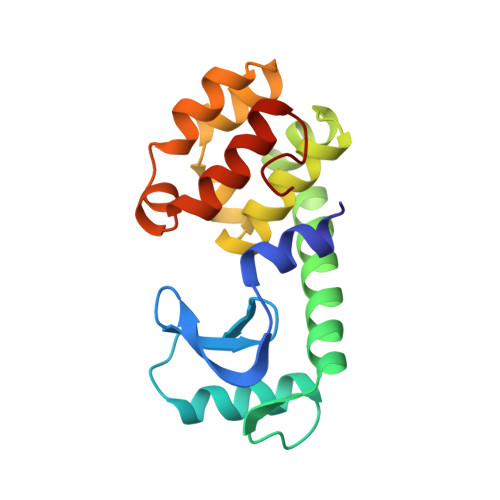
1L55, 1L57, 1L59, 1L61, 1L62, 1L63 - PubMed Abstract:
It was shown previously that the introduction of a negatively charged amino acid at the N-terminus of an alpha-helix could increase the thermostability of phage T4 lysozyme via an electrostatic interaction with the "helix dipole" [Nicholson, H., Becktel, W. J., & Matthews, B. W. (1988) Nature 336, 651-656]. The prior report focused on the two stabilizing substitutions Ser 38----Asp (S38D) and Asn 144----Asp (N144D). Two additional examples of stabilizing mutants, T109D and N116D, are presented here. Both show the pH-dependent increase in thermal stability expected for the interaction of an aspartic acid with an alpha-helix dipole. Control mutants were also constructed to further characterize the nature of the interaction with the alpha-helix dipole. High-resolution crystal structure analysis was used to determine the nature of the interaction of the substituted amino acids with the end of the alpha-helix in both the primary and the control mutants. Control mutant S38N has stability essentially the same as that of wild-type lysozyme but hydrogen bonding similar to that of the stabilizing mutant S38D. This confirms that it is the electrostatic interaction between Asp 38 and the helix dipole, rather than a change in hydrogen-bonding geometry, that gives enhanced stability. Structural and thermodynamic analysis of mutant T109N provide a similar control for the stabilizing replacement T109D. In the case of mutant N116D, there was concern that the enhanced stability might be due to a favorable salt-bridge interaction between the introduced aspartate and Arg 119, rather than an interaction with the alpha-helix dipole. The additivity of the stabilities of N116D and R119M seen in the double mutant N116D/R119M indicates that favorable interactions are largely independent of residue 119. As a further control, Asp 92, a presumed helix-stabilizing residue in wild-type lysozyme, was replaced with Asn. This decreased the stability of the protein in the manner expected for the loss of a favorable helix dipole interaction. In total, five mutations have been identified that increase the thermostability of T4 lysozyme and appear to do so by favorable interactions with alpha-helix dipoles. As measured by the pH dependence of stability, the strength of the electrostatic interaction between the charged groups studied here and the helix dipole ranges from 0.6 to 1.3 kcal/mol in 150 mM KCl. In the case of mutants S38D and N144H, NMR titration was used to measure the pKa's of Asp 38 and His 144 in the folded structures.(ABSTRACT TRUNCATED AT 400 WORDS)
Organizational Affiliation:
Institute of Molecular Biology, Howard Hughes Medical Institute, Eugene, Oregon.