Contributions of left-handed helical residues to the structure and stability of bacteriophage T4 lysozyme.
Nicholson, H., Soderlind, E., Tronrud, D.E., Matthews, B.W.(1989) J Mol Biol 210: 181-193
- PubMed: 2511328
- DOI: https://doi.org/10.1016/0022-2836(89)90299-4
- Primary Citation of Related Structures:
1L21, 1L22, 1L33 - PubMed Abstract:
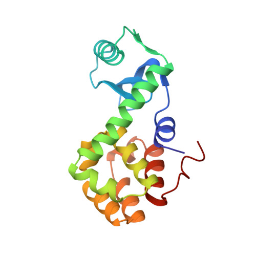
Non-glycine residues in proteins are rarely observed to have "left-handed helical" conformations. For glycine, however, this conformation is common. To determine the contributions of left-handed helical residues to the stability of a protein, two such residues in phage T4 lysozyme, Asn55 and Lys124, were replaced with glycine. The mutant proteins fold normally and are fully active, showing that left-handed non-glycine residues, although rare, do not have an indispensable role in the folding of the protein or in its activity. The thermodynamic stability of the Lys124 to Gly variant is essentially identical with that of wild-type lysozyme. The Asn55 to Gly mutant protein is marginally less stable (0.5 kcal/mol). These results indicate that the conformational energy of a glycine and a non-glycine residue in the left-handed helical conformation are very similar. This is consistent with some theoretical energy distributions, but is inconsistent with others, which suggest that replacements of the sort described here might increase the stability of the protein by up to 5 kcal/mol. Crystallographic analysis of the mutant proteins shows that the backbone conformation of the Lys124 to Gly variant is essentially identical with that of the wild-type structure. In the case of the Asn55 to Gly replacement, however, the (phi, psi) values of residue 55 change by about 20 degrees. This suggests that the energy minimum for left-handed glycine residues is not the same as that for non-glycine residues. This is strongly indicated also by a survey of accurately determined protein crystal structures, which suggests that the energy minimum for left-handed glycine residues is near (phi = 90 degrees, psi = 0 degrees), whereas that for non-glycine residues is close to (phi = 60 degrees, psi = 30 degrees). This apparent energy minimum for glycine is not clearly predicted by any of the theoretical (phi, psi) energy contour maps.
Organizational Affiliation:
Institute of Molecular Biology, University of Oregon, Eugene 97403.