1JQU

Are Carboxy Terminii of Helices Coded by the Local Sequence or by Tertiary Structure Contacts


Experimental Data Snapshot

  • Method: X-RAY DIFFRACTION
  • Resolution: 2.60 Å
  • R-Value Free: 0.316 
  • R-Value Work: 0.229 
  • R-Value Observed: 0.241 

wwPDB Validation   3D Report Full Report


This is version 1.7 of the entry. See complete history


Literature

A test of proposed rules for helix capping: Implications for protein design

Sagermann, M.Martensson, L.-G.Baase, W.A.Matthews, B.W.

(2002) Protein Sci 11: 516-521

  • DOI: https://doi.org/10.1110/ps.39802
  • Primary Citation of Related Structures:  
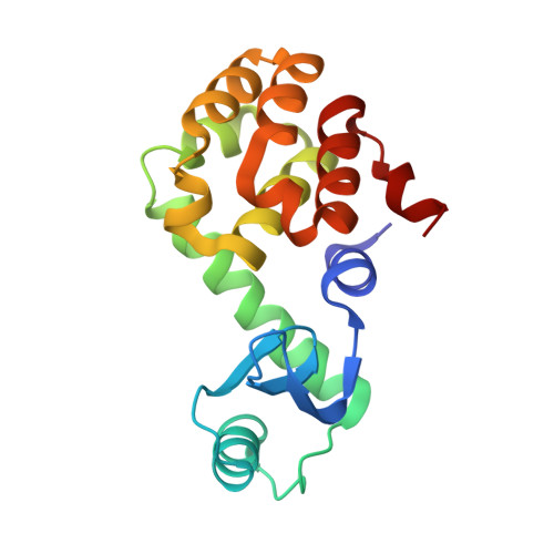
    1JQU, 1LLH

  • PubMed Abstract: 

    alpha-helices within proteins are often terminated (capped) by distinctive configurations of the polypeptide chain. Two common arrangements are the Schellman motif and the alternative alpha(L) motif. Rose and coworkers developed stereochemical rules to identify the locations of such motifs in proteins of unknown structure based only on their amino acid sequences. To check the effectiveness of these rules, they made specific predictions regarding the structural and thermodynamic consequences of certain mutations in T4 lysozyme. We have constructed these mutants and show here that they have neither the structure nor the stability that was predicted. The results show the complexity of the protein-folding problem. Comparison of known protein structures may show that a characteristic sequence of amino acids (a sequence motif) corresponds to a conserved structural motif. In any particular protein, however, changes in other parts of the sequence may result in a different conformation. The structure is determined by sequence as a whole, not by parts considered in isolation.


  • Organizational Affiliation

    Institute of Molecular Biology, Howard Hughes Medical Institute and Department of Physics, University of Oregon, Eugene, Oregon 97403-1229, USA.


Macromolecules
Find similar proteins by:  (by identity cutoff)  |  3D Structure
Entity ID: 1
MoleculeChains Sequence LengthOrganismDetailsImage
Lysozyme
A, B, C, D
164Tequatrovirus T4Mutation(s): 3 
Gene Names: E
EC: 3.2.1.17
UniProt
Find proteins for P00720 (Enterobacteria phage T4)
Explore P00720 
Go to UniProtKB:  P00720
Entity Groups  
Sequence Clusters30% Identity50% Identity70% Identity90% Identity95% Identity100% Identity
UniProt GroupP00720
Sequence Annotations
Expand
  • Reference Sequence
Experimental Data & Validation

Experimental Data

  • Method: X-RAY DIFFRACTION
  • Resolution: 2.60 Å
  • R-Value Free: 0.316 
  • R-Value Work: 0.229 
  • R-Value Observed: 0.241 
  • Space Group: P 21 21 21
Unit Cell:
Length ( Å )Angle ( ˚ )
a = 74.3α = 90
b = 77.2β = 90
c = 138.2γ = 90
Software Package:
Software NamePurpose
TNTrefinement
CNSrefinement
CNSphasing
TNTphasing

Structure Validation

View Full Validation Report


Entry History 

Deposition Data

Revision History  (Full details and data files)

  • Version 1.0: 2002-03-06
    Type: Initial release
  • Version 1.1: 2008-04-27
    Changes: Version format compliance
  • Version 1.2: 2011-07-13
    Changes: Version format compliance
  • Version 1.3: 2014-03-26
    Changes: Database references
  • Version 1.4: 2017-10-04
    Changes: Refinement description
  • Version 1.5: 2019-07-24
    Changes: Data collection, Refinement description
  • Version 1.6: 2021-10-27
    Changes: Database references
  • Version 1.7: 2023-08-16
    Changes: Data collection, Refinement description