1IQQ

Crystal Structure of Japanese pear S3-RNase


Experimental Data Snapshot

  • Method: X-RAY DIFFRACTION
  • Resolution: 1.50 Å
  • R-Value Free: 0.202 
  • R-Value Work: 0.172 
  • R-Value Observed: 0.172 

wwPDB Validation   3D Report Full Report


This is version 2.1 of the entry. See complete history


Literature

Crystal structure at 1.5-A resolution of Pyrus pyrifolia pistil ribonuclease responsible for gametophytic self-incompatibility.

Matsuura, T.Sakai, H.Unno, M.Ida, K.Sato, M.Sakiyama, F.Norioka, S.

(2001) J Biol Chem 276: 45261-45269

  • DOI: https://doi.org/10.1074/jbc.M107617200
  • Primary Citation of Related Structures:  
    1IQQ

  • PubMed Abstract: 

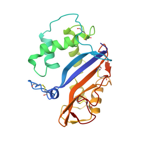
    The crystal structure of the Pyrus pyrifolia pistil ribonuclease (S(3)-RNase) responsible for gametophytic self-incompatibility was determined at 1.5-A resolution. It consists of eight helices and seven beta-strands, and its folding topology is typical of RNase T(2) family enzymes. Based on a structural comparison of S(3)-RNase with RNase Rh, a fungal RNase T(2) family enzyme, the active site residues of S(3)-RNase assigned were His(33) and His(88) as catalysts and Glu(84) and Lys(87) as stabilizers of an intermediate in the transition state. Moreover, amino acid residues that constitute substrate binding sites of the two RNases could be superimposed geometrically. A hypervariable (HV) region that has an S-allele-specific sequence comprises a long loop and short alpha-helix. This region is far from the active site cleft, exposed on the molecule's surface, and positively charged. Four positively selected (PS) regions, in which the number of nonsynonymous substitutions exceeds that of synonymous ones, are located on either side of the active site cleft, and accessible to solvent. These structural features suggest that the HV or PS regions may interact with a pollen S-gene product(s) to recognize self and non-self pollen.


  • Organizational Affiliation

    Institute for Protein Research, Osaka University, Suita, Osaka 565-0871, Japan.


Macromolecules
Find similar proteins by:  (by identity cutoff)  |  3D Structure
Entity ID: 1
MoleculeChains Sequence LengthOrganismDetailsImage
S3-RNase200Pyrus pyrifoliaMutation(s): 0 
EC: 3.1.27.1
UniProt
Find proteins for O80323 (Pyrus pyrifolia)
Explore O80323 
Go to UniProtKB:  O80323
Entity Groups  
Sequence Clusters30% Identity50% Identity70% Identity90% Identity95% Identity100% Identity
UniProt GroupO80323
Sequence Annotations
Expand
  • Reference Sequence
Oligosaccharides

Help

Entity ID: 2
MoleculeChains Length2D Diagram Glycosylation3D Interactions
beta-D-xylopyranose-(1-2)-[alpha-D-mannopyranose-(1-3)]beta-D-mannopyranose-(1-4)-2-acetamido-2-deoxy-beta-D-glucopyranose-(1-4)-2-acetamido-2-deoxy-beta-D-glucopyranose
B
5N-Glycosylation
Glycosylation Resources
GlyTouCan:  G71469XA
GlyCosmos:  G71469XA
GlyGen:  G71469XA
Experimental Data & Validation

Experimental Data

  • Method: X-RAY DIFFRACTION
  • Resolution: 1.50 Å
  • R-Value Free: 0.202 
  • R-Value Work: 0.172 
  • R-Value Observed: 0.172 
  • Space Group: P 1 21 1
Unit Cell:
Length ( Å )Angle ( ˚ )
a = 45.65α = 90
b = 52.59β = 106.45
c = 47.57γ = 90
Software Package:
Software NamePurpose
DENZOdata reduction
SCALEPACKdata scaling
SHARPphasing
CNSrefinement

Structure Validation

View Full Validation Report


Entry History 

Deposition Data

Revision History  (Full details and data files)

  • Version 1.0: 2001-11-07
    Type: Initial release
  • Version 1.1: 2008-04-27
    Changes: Version format compliance
  • Version 1.2: 2011-07-13
    Changes: Non-polymer description, Version format compliance
  • Version 2.0: 2020-07-29
    Type: Remediation
    Reason: Carbohydrate remediation
    Changes: Atomic model, Data collection, Derived calculations, Structure summary
  • Version 2.1: 2023-12-27
    Changes: Data collection, Database references, Derived calculations, Structure summary