1HNF

CRYSTAL STRUCTURE OF THE EXTRACELLULAR REGION OF THE HUMAN CELL ADHESION MOLECULE CD2 AT 2.5 ANGSTROMS RESOLUTION


Experimental Data Snapshot

  • Method: X-RAY DIFFRACTION
  • Resolution: 2.50 Å
  • R-Value Work: 0.193 
  • R-Value Observed: 0.193 

wwPDB Validation   3D Report Full Report


This is version 1.3 of the entry. See complete history


Literature

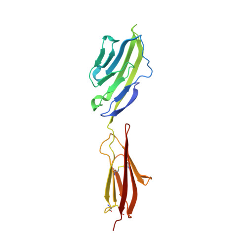
Crystal structure of the extracellular region of the human cell adhesion molecule CD2 at 2.5 A resolution.

Bodian, D.L.Jones, E.Y.Harlos, K.Stuart, D.I.Davis, S.J.

(1994) Structure 2: 755-766

  • DOI: https://doi.org/10.1016/s0969-2126(94)00076-x
  • Primary Citation of Related Structures:  
    1HNF

  • PubMed Abstract: 

    The T-lymphocyte antigen CD2 is an adhesion molecule implicated in immune responses in vivo. The extracellular regions of the human and rat homologues of CD2 share only 45% sequence identity and bind different protein ligands. Comparison of the human and rat soluble CD2 (sCD2) structures should provide insights into the structural basis of cell surface recognition. We therefore determined the crystal structure of a form of human sCD2 with single N-acetylglucosamine residues at each glycosylation site to 2.5 A resolution with an R-factor of 19.3%. It is composed of two immunoglobulin superfamily domains similar to those of rat sCD2, but the relative orientation of the domains in the two homologues differs by up to 20 degrees. An interaction involving the flat, highly charged, ligand binding GFCC'C" faces of crystallographically related human sCD2 molecules duplicates, in a different lattice, that observed in the rat sCD2 crystals. Intramolecular flexibility appears to be a conserved feature of CD2. The head-to-head interaction between molecules represents a general model for interactions between adhesion molecules of this structural class. Ligand specificity may be influenced by the distribution of charged residues on the binding face.


  • Organizational Affiliation

    Laboratory of Molecular Biophysics, Oxford, UK.


Macromolecules
Find similar proteins by:  (by identity cutoff)  |  3D Structure
Entity ID: 1
MoleculeChains Sequence LengthOrganismDetailsImage
CD2182Homo sapiensMutation(s): 0 
UniProt & NIH Common Fund Data Resources
Find proteins for P06729 (Homo sapiens)
Explore P06729 
Go to UniProtKB:  P06729
PHAROS:  P06729
GTEx:  ENSG00000116824 
Entity Groups  
Sequence Clusters30% Identity50% Identity70% Identity90% Identity95% Identity100% Identity
UniProt GroupP06729
Sequence Annotations
Expand
  • Reference Sequence
Small Molecules
Experimental Data & Validation

Experimental Data

  • Method: X-RAY DIFFRACTION
  • Resolution: 2.50 Å
  • R-Value Work: 0.193 
  • R-Value Observed: 0.193 
  • Space Group: P 31 2 1
Unit Cell:
Length ( Å )Angle ( ˚ )
a = 88.3α = 90
b = 88.3β = 90
c = 51.3γ = 120
Software Package:
Software NamePurpose
X-PLORmodel building
X-PLORrefinement
X-PLORphasing

Structure Validation

View Full Validation Report


Entry History 

Deposition Data

Revision History  (Full details and data files)

  • Version 1.0: 1995-02-07
    Type: Initial release
  • Version 1.1: 2008-03-24
    Changes: Version format compliance
  • Version 1.2: 2011-07-13
    Changes: Non-polymer description, Version format compliance
  • Version 1.3: 2020-07-29
    Type: Remediation
    Reason: Carbohydrate remediation
    Changes: Data collection, Derived calculations, Other, Structure summary