1H2P

Human CD55 domains 3 & 4


Experimental Data Snapshot

  • Method: X-RAY DIFFRACTION
  • Resolution: 2.80 Å
  • R-Value Free: 0.304 
  • R-Value Work: 0.258 
  • R-Value Observed: 0.258 

wwPDB Validation   3D Report Full Report


This is version 1.3 of the entry. See complete history


Literature

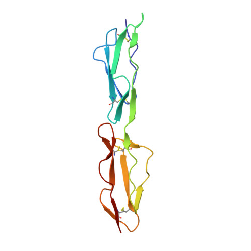
Mapping Cd55 Function. The Structure of Two Pathogen-Binding Domains at 1.7 A

Williams, P.Chaudhry, Y.Goodfellow, I.Billington, J.Powell, R.Spiller, O.Evans, D.J.Lea, S.M.

(2003) J Biol Chem 278: 10691

  • DOI: https://doi.org/10.1074/jbc.M212561200
  • Primary Citation of Related Structures:  
    1H03, 1H04, 1H2P, 1H2Q, 1UOT

  • PubMed Abstract: 

    Decay-accelerating factor (CD55), a regulator of the alternative and classical pathways of complement activation, is expressed on all serum-exposed cells. It is used by pathogens, including many enteroviruses and uropathogenic Escherichia coli, as a receptor prior to infection. We describe the x-ray structure of a pathogen-binding fragment of human CD55 at 1.7 A resolution containing two of the three domains required for regulation of human complement. We have used mutagenesis to map biological functions onto the molecule; decay-accelerating activity maps to a single face of the molecule, whereas bacterial and viral pathogens recognize a variety of different sites on CD55.


  • Organizational Affiliation

    Laboratory of Molecular Biophysics, Department of Biochemistry, University of Oxford, United Kingdom.


Macromolecules
Find similar proteins by:  (by identity cutoff)  |  3D Structure
Entity ID: 1
MoleculeChains Sequence LengthOrganismDetailsImage
COMPLEMENT DECAY-ACCELERATING FACTORA [auth P]125Homo sapiensMutation(s): 0 
UniProt & NIH Common Fund Data Resources
Find proteins for P08174 (Homo sapiens)
Explore P08174 
Go to UniProtKB:  P08174
PHAROS:  P08174
GTEx:  ENSG00000196352 
Entity Groups  
Sequence Clusters30% Identity50% Identity70% Identity90% Identity95% Identity100% Identity
UniProt GroupP08174
Sequence Annotations
Expand
  • Reference Sequence
Experimental Data & Validation

Experimental Data

  • Method: X-RAY DIFFRACTION
  • Resolution: 2.80 Å
  • R-Value Free: 0.304 
  • R-Value Work: 0.258 
  • R-Value Observed: 0.258 
  • Space Group: C 1 2 1
Unit Cell:
Length ( Å )Angle ( ˚ )
a = 126.9α = 90
b = 20.7β = 99.4
c = 41.8γ = 90
Software Package:
Software NamePurpose
CNSrefinement
DENZOdata reduction
SCALEPACKdata scaling
EPMRphasing

Structure Validation

View Full Validation Report


Entry History 

Deposition Data

Revision History  (Full details and data files)

  • Version 1.0: 2003-03-20
    Type: Initial release
  • Version 1.1: 2011-05-08
    Changes: Version format compliance
  • Version 1.2: 2011-07-13
    Changes: Version format compliance
  • Version 1.3: 2023-12-13
    Changes: Data collection, Database references, Refinement description