1GGU

HUMAN FACTOR XIII WITH CALCIUM BOUND IN THE ION SITE


Experimental Data Snapshot

  • Method: X-RAY DIFFRACTION
  • Resolution: 2.10 Å
  • R-Value Free: 0.313 
  • R-Value Work: 0.227 
  • R-Value Observed: 0.227 

wwPDB Validation   3D Report Full Report


This is version 1.3 of the entry. See complete history


Literature

Identification of the calcium binding site and a novel ytterbium site in blood coagulation factor XIII by x-ray crystallography.

Fox, B.A.Yee, V.C.Pedersen, L.C.Le Trong, I.Bishop, P.D.Stenkamp, R.E.Teller, D.C.

(1999) J Biol Chem 274: 4917-4923

  • DOI: https://doi.org/10.1074/jbc.274.8.4917
  • Primary Citation of Related Structures:  
    1GGU, 1GGY, 1QRK

  • PubMed Abstract: 

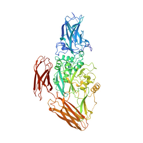
    The presence or absence of calcium determines the activation, activity, oligomerization, and stability of blood coagulation factor XIII. To explore these observed effects, we have determined the x-ray crystal structure of recombinant factor XIII A2 in the presence of calcium, strontium, and ytterbium. The main calcium binding site within each monomer involves the main chain oxygen atom of Ala-457, and also the side chains from residues Asn-436, Asp-438, Glu-485, and Glu-490. Calcium and strontium bind in the same location, while ytterbium binds several angstroms removed. A novel ytterbium binding site is also found at the dimer two-fold axis, near residues Asp-270 and Glu-272, and this site may be related to the reported inhibition by lanthanide metals (Achyuthan, K. E., Mary, A., and Greenberg, C. S. (1989) Biochem. J. 257, 331-338). The overall structure of ion-bound factor XIII is very similar to the previously determined crystal structures of factor XIII zymogen, likely due to the constraints of this monoclinic crystal form. We have merged the three independent sets of water molecules in the structures to determine which water molecules are conserved and possibly structurally significant.


  • Organizational Affiliation

    Department of Biochemistry, University of Washington, Seattle, Washington 98195, USA.


Macromolecules
Find similar proteins by:  (by identity cutoff)  |  3D Structure
Entity ID: 1
MoleculeChains Sequence LengthOrganismDetailsImage
PROTEIN (COAGULATION FACTOR XIII)
A, B
731Homo sapiensMutation(s): 0 
EC: 2.3.2.13
UniProt & NIH Common Fund Data Resources
Find proteins for P00488 (Homo sapiens)
Explore P00488 
Go to UniProtKB:  P00488
PHAROS:  P00488
GTEx:  ENSG00000124491 
Entity Groups  
Sequence Clusters30% Identity50% Identity70% Identity90% Identity95% Identity100% Identity
UniProt GroupP00488
Sequence Annotations
Expand
  • Reference Sequence
Small Molecules
Ligands 1 Unique
IDChains Name / Formula / InChI Key2D Diagram3D Interactions
CA
Query on CA

Download Ideal Coordinates CCD File 
C [auth A],
D [auth B]
CALCIUM ION
Ca
BHPQYMZQTOCNFJ-UHFFFAOYSA-N
Experimental Data & Validation

Experimental Data

  • Method: X-RAY DIFFRACTION
  • Resolution: 2.10 Å
  • R-Value Free: 0.313 
  • R-Value Work: 0.227 
  • R-Value Observed: 0.227 
  • Space Group: P 1 21 1
Unit Cell:
Length ( Å )Angle ( ˚ )
a = 100.172α = 90
b = 70.764β = 106.11
c = 133.822γ = 90
Software Package:
Software NamePurpose
X-PLORmodel building
X-PLORrefinement
DENZOdata reduction
SCALEPACKdata scaling
X-PLORphasing

Structure Validation

View Full Validation Report


Entry History 

Deposition Data

Revision History  (Full details and data files)

  • Version 1.0: 1999-09-16
    Type: Initial release
  • Version 1.1: 2007-10-16
    Changes: Version format compliance
  • Version 1.2: 2011-07-13
    Changes: Version format compliance
  • Version 1.3: 2023-08-09
    Changes: Data collection, Database references, Derived calculations, Refinement description