1G7B

1.3 A STRUCTURE OF T3R3 HUMAN INSULIN AT 100 K


Experimental Data Snapshot

  • Method: X-RAY DIFFRACTION
  • Resolution: 1.30 Å
  • R-Value Free: 0.204 
  • R-Value Work: 0.176 

wwPDB Validation   3D Report Full Report


This is version 1.4 of the entry. See complete history


Literature

Phase changes in T(3)R(3)(f) human insulin: temperature or pressure induced?

Smith, G.D.Pangborn, W.A.Blessing, R.H.

(2001) Acta Crystallogr D Biol Crystallogr 57: 1091-1100

  • DOI: https://doi.org/10.1107/s0907444901007685
  • Primary Citation of Related Structures:  
    1G7A, 1G7B

  • PubMed Abstract: 

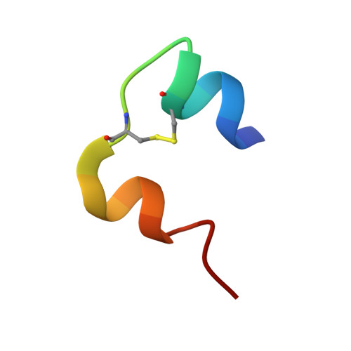
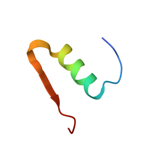
    The structure of T(3)R(3) hexameric human insulin has been determined at 100 K from two different crystals at 1.2 and 1.3 A resolution and refined to residuals of 0.169 and 0.176, respectively. Owing to a phase change, the c axis is double its room-temperature value and the asymmetric unit contains two independent TR(f) insulin dimers. Compared with the orientation in the room-temperature structure, one dimer undergoes a rotation about the c axis of -5 degrees, while the second is rotated +4 degrees. A superposition of the backbone atoms of the two independent dimers shows that the C(alpha) atoms of five residues within the R(f)-state monomers are displaced by more than 1.0 A; smaller displacements are observed for the T-state monomers. Four zinc ions lie on the crystallographic threefold axis and each forms bonds to three symmetry-related HisB10 N(varepsilon2) atoms from the T- and R(f)-state trimers. While three of the zinc ions are tetrahedrally coordinated with a chloride ion completing the coordination sphere, mixed tetrahedral/octahedral coordination is observed for one of the T-state zinc ions. The three symmetry-related "phenolic binding sites" in one hexamer contain water molecules and a glycerol molecule, but the same sites in the second hexamer are occupied by a zinc ion coordinated to an alternate conformation of HisB10, a symmetry-related HisB5 and two chloride ions. Two additional and partially occupied zinc ion sites are observed at the interface between the two independent dimers. One zinc ion is coordinated by a T-state HisB5 of one dimer, an R-state HisB5 of the second dimer and two water molecules; the second zinc ion is coordinated by an alternate side-chain conformation of the T-state HisB5 and three water molecules. The carboxyl group of one GluB13 side chain, which exists in two discrete conformations, appears to be protonated, because short contacts exist to a second carboxyl group or to a carbonyl O atom.


  • Organizational Affiliation

    Hauptman-Woodward Medical Research Institute, 73 High Street, Buffalo, NY 14203, USA. smith@hwi.buffalo.edu


Macromolecules

Find similar proteins by:  Sequence   |   3D Structure  

Entity ID: 1
MoleculeChains Sequence LengthOrganismDetailsImage
INSULIN A-CHAIN
A, C, E, G
21N/AMutation(s): 0 
UniProt & NIH Common Fund Data Resources
Find proteins for P01308 (Homo sapiens)
Explore P01308 
Go to UniProtKB:  P01308
PHAROS:  P01308
GTEx:  ENSG00000254647 
Entity Groups  
Sequence Clusters30% Identity50% Identity70% Identity90% Identity95% Identity100% Identity
UniProt GroupP01308
Sequence Annotations
Expand
  • Reference Sequence
Find similar proteins by:  (by identity cutoff)  |  3D Structure
Entity ID: 2
MoleculeChains Sequence LengthOrganismDetailsImage
INSULIN B-CHAIN
B, D, F, H
30N/AMutation(s): 0 
UniProt & NIH Common Fund Data Resources
Find proteins for P01308 (Homo sapiens)
Explore P01308 
Go to UniProtKB:  P01308
PHAROS:  P01308
GTEx:  ENSG00000254647 
Entity Groups  
Sequence Clusters30% Identity50% Identity70% Identity90% Identity95% Identity100% Identity
UniProt GroupP01308
Sequence Annotations
Expand
  • Reference Sequence
Small Molecules
Ligands 4 Unique
IDChains Name / Formula / InChI Key2D Diagram3D Interactions
GOL
Query on GOL

Download Ideal Coordinates CCD File 
U [auth G]GLYCEROL
C3 H8 O3
PEDCQBHIVMGVHV-UHFFFAOYSA-N
ZN
Query on ZN

Download Ideal Coordinates CCD File 
I [auth B]
J [auth B]
K [auth B]
M [auth D]
N [auth D]
I [auth B],
J [auth B],
K [auth B],
M [auth D],
N [auth D],
S [auth F],
W [auth H]
ZINC ION
Zn
PTFCDOFLOPIGGS-UHFFFAOYSA-N
ACN
Query on ACN

Download Ideal Coordinates CCD File 
V [auth G]ACETONE
C3 H6 O
CSCPPACGZOOCGX-UHFFFAOYSA-N
CL
Query on CL

Download Ideal Coordinates CCD File 
L [auth B]
O [auth D]
P [auth D]
Q [auth D]
R [auth E]
L [auth B],
O [auth D],
P [auth D],
Q [auth D],
R [auth E],
T [auth F],
X [auth H]
CHLORIDE ION
Cl
VEXZGXHMUGYJMC-UHFFFAOYSA-M
Experimental Data & Validation

Experimental Data

  • Method: X-RAY DIFFRACTION
  • Resolution: 1.30 Å
  • R-Value Free: 0.204 
  • R-Value Work: 0.176 
  • Space Group: H 3
Unit Cell:
Length ( Å )Angle ( ˚ )
a = 80.05α = 90
b = 80.05β = 90
c = 71.459γ = 120
Software Package:
Software NamePurpose
DENZOdata reduction
SCALEPACKdata scaling
CNSrefinement
CNSphasing

Structure Validation

View Full Validation Report


Entry History 

Deposition Data

Revision History  (Full details and data files)

  • Version 1.0: 2001-08-03
    Type: Initial release
  • Version 1.1: 2008-04-27
    Changes: Version format compliance
  • Version 1.2: 2011-07-13
    Changes: Non-polymer description, Version format compliance
  • Version 1.3: 2019-11-20
    Changes: Derived calculations
  • Version 1.4: 2023-08-09
    Changes: Data collection, Database references, Derived calculations, Refinement description