1ESO

MONOMERIC CU,ZN SUPEROXIDE DISMUTASE FROM ESCHERICHIA COLI


Experimental Data Snapshot

  • Method: X-RAY DIFFRACTION
  • Resolution: 2.00 Å
  • R-Value Free: 0.260 
  • R-Value Work: 0.168 

wwPDB Validation   3D Report Full Report


This is version 1.3 of the entry. See complete history


Literature

Unique structural features of the monomeric Cu,Zn superoxide dismutase from Escherichia coli, revealed by X-ray crystallography.

Pesce, A.Capasso, C.Battistoni, A.Folcarelli, S.Rotilio, G.Desideri, A.Bolognesi, M.

(1997) J Mol Biol 274: 408-420

  • DOI: https://doi.org/10.1006/jmbi.1997.1400
  • Primary Citation of Related Structures:  
    1ESO

  • PubMed Abstract: 

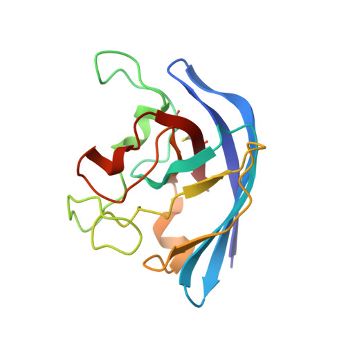
    The first three-dimensional structure of a functional monomeric Cu, Zn superoxide dismutase (from Escherichia coli, E_SOD) is reported at 2.0 A resolution (R-factor=16.8%). Compared to the homologous eukaryotic enzymes, E_SOD displays a perturbed antiparallel beta-barrel structure. The most striking structural features observed include extended amino acid insertions in the surface 1, 2-loop and S-S subloop, modification of the disulfide bridge connection, and loss of functional electrostatic residues, suggesting a modified control of substrate steering toward the catalytic center. The active site Cu2+ displays a distorted coordination sphere due to an unusually long bond to the metal-bridging residue His61. Inspection of the crystal packing does not show regions of extended contact indicative of a dimeric assembly. The molecular surface region involved in subunit dimerization in eukaryotic superoxide dismutases is structurally altered in E_SOD and displays a net polar nature.


  • Organizational Affiliation

    Dipartimento di Fisica and INFM, Universita' di Genova, Largo Rosanna Benzi, 10, 16132 Genova, Italy.


Macromolecules
Find similar proteins by:  (by identity cutoff)  |  3D Structure
Entity ID: 1
MoleculeChains Sequence LengthOrganismDetailsImage
CU, ZN SUPEROXIDE DISMUTASE154Escherichia coliMutation(s): 0 
Gene Names: SODC
EC: 1.15.1.1
UniProt
Find proteins for P0AGD1 (Escherichia coli (strain K12))
Explore P0AGD1 
Go to UniProtKB:  P0AGD1
Entity Groups  
Sequence Clusters30% Identity50% Identity70% Identity90% Identity95% Identity100% Identity
UniProt GroupP0AGD1
Sequence Annotations
Expand
  • Reference Sequence
Experimental Data & Validation

Experimental Data

  • Method: X-RAY DIFFRACTION
  • Resolution: 2.00 Å
  • R-Value Free: 0.260 
  • R-Value Work: 0.168 
  • Space Group: P 1 21 1
Unit Cell:
Length ( Å )Angle ( ˚ )
a = 33α = 90
b = 52.4β = 111.2
c = 43.2γ = 90
Software Package:
Software NamePurpose
MOSFLMdata reduction
ROTAVATAdata reduction
Agrovatadata reduction
AMoREphasing
TNTrefinement
CCP4data scaling
ROTAVATAdata scaling

Structure Validation

View Full Validation Report


Entry History 

Deposition Data

Revision History  (Full details and data files)

  • Version 1.0: 1998-07-01
    Type: Initial release
  • Version 1.1: 2008-03-24
    Changes: Version format compliance
  • Version 1.2: 2011-07-13
    Changes: Version format compliance
  • Version 1.3: 2024-04-03
    Changes: Data collection, Database references, Derived calculations, Refinement description