1EFD

PERIPLASMIC FERRIC SIDEROPHORE BINDING PROTEIN FHUD COMPLEXED WITH GALLICHROME


Experimental Data Snapshot

  • Method: X-RAY DIFFRACTION
  • Resolution: 1.90 Å
  • R-Value Free: 0.250 
  • R-Value Work: 0.218 

wwPDB Validation   3D Report Full Report


This is version 1.3 of the entry. See complete history


Literature

The structure of the ferric siderophore binding protein FhuD complexed with gallichrome.

Clarke, T.E.Ku, S.Y.Dougan, D.R.Vogel, H.J.Tari, L.W.

(2000) Nat Struct Biol 7: 287-291

  • DOI: https://doi.org/10.1038/74048
  • Primary Citation of Related Structures:  
    1EFD

  • PubMed Abstract: 

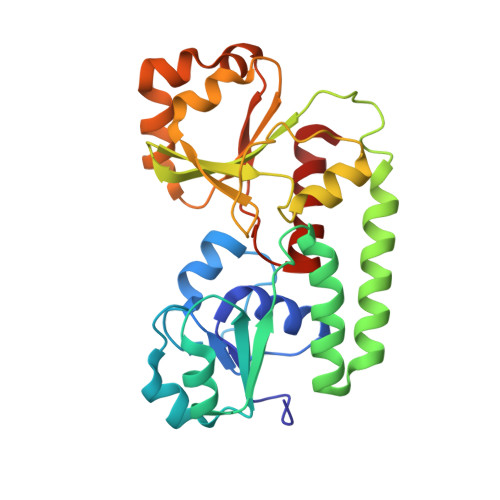
    Siderophore binding proteins play a key role in the uptake of iron in many gram-positive and gram-negative bacteria. FhuD is a soluble periplasmic binding protein that transports ferrichrome and other hydroxamate siderophores. The crystal structure of FhuD from Escherichia coli in complex with the ferrichrome homolog gallichrome has been determined at 1.9 ¿ resolution, the first structure of a periplasmic binding protein involved in the uptake of siderophores. Gallichrome is held in a shallow pocket lined with aromatic groups; Arg and Tyr side chains interact directly with the hydroxamate moieties of the siderophore. FhuD possesses a novel fold, suggesting that its mechanisms of ligand binding and release are different from other structurally characterized periplasmic ligand binding proteins.


  • Organizational Affiliation

    Department of Biological Sciences, University of Calgary, 2500 University Drive N.W., Calgary, Alberta, Canada T2N 1N4.


Macromolecules
Find similar proteins by:  (by identity cutoff)  |  3D Structure
Entity ID: 1
MoleculeChains Sequence LengthOrganismDetailsImage
FERRICHROME-BINDING PERIPLASMIC PROTEINA [auth N]266Escherichia coliMutation(s): 0 
UniProt
Find proteins for P07822 (Escherichia coli (strain K12))
Explore P07822 
Go to UniProtKB:  P07822
Entity Groups  
Sequence Clusters30% Identity50% Identity70% Identity90% Identity95% Identity100% Identity
UniProt GroupP07822
Sequence Annotations
Expand
  • Reference Sequence
Small Molecules
Ligands 1 Unique
IDChains Name / Formula / InChI Key2D Diagram3D Interactions
GCR
Query on GCR

Download Ideal Coordinates CCD File 
B [auth N]GALLICHROME
C28 H44 Ga N9 O13
BEDMDXBTNSVNGS-GSJIPYROSA-N
Experimental Data & Validation

Experimental Data

  • Method: X-RAY DIFFRACTION
  • Resolution: 1.90 Å
  • R-Value Free: 0.250 
  • R-Value Work: 0.218 
  • Space Group: P 63
Unit Cell:
Length ( Å )Angle ( ˚ )
a = 86.54α = 90
b = 86.54β = 90
c = 91.77γ = 120
Software Package:
Software NamePurpose
CNSrefinement
DENZOdata reduction
SCALEPACKdata scaling
CNSphasing

Structure Validation

View Full Validation Report


Entry History 

Deposition Data

Revision History  (Full details and data files)

  • Version 1.0: 2000-04-05
    Type: Initial release
  • Version 1.1: 2008-04-27
    Changes: Version format compliance
  • Version 1.2: 2011-07-13
    Changes: Version format compliance
  • Version 1.3: 2024-02-07
    Changes: Data collection, Database references, Derived calculations