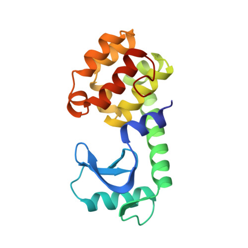
Determination of alpha-helix propensity within the context of a folded protein. Sites 44 and 131 in bacteriophage T4 lysozyme.
Blaber, M., Zhang, X.J., Lindstrom, J.D., Pepiot, S.D., Baase, W.A., Matthews, B.W.(1994) J Mol Biol 235: 600-624
- PubMed: 8289284
- DOI: https://doi.org/10.1006/jmbi.1994.1016
- Primary Citation of Related Structures:
1DYA, 1DYB, 1DYC, 1DYD, 1DYE, 1DYF, 1DYG, 3L64 - PubMed Abstract:
To determine the effects of different amino acids on the structure and stability of an alpha-helix in the context of a globular protein, all 19 naturally-occurring amino acids were substituted for Ser44 in phage T4 lysozyme. A more restricted set of nine replacements was also made for Val131. Ser44 and Val131 are two of a very limited number of possible sites in T4 lysozyme that are well within alpha-helices, are solvent-exposed and relatively free of interactions with neighboring residues, and are not involved in crystal contacts. High resolution structures for the majority of the mutants, some of which crystallized non-isomorphously with wild-type, were determined. With the exception of proline, the amino acid substitutions caused little if any perturbation of the alpha-helix backbone. Also the beta-branched residues Thr, Val and Ile show no indication of either side-chain or backbone distortion. Therefore, other than proline, there is no evidence that differences in helix propensities are associated with different amounts of strain introduced into the helix. For reference, and also to allow estimates of side-chain entropy, a survey was made of side-chain conformations in 100 well-refined protein structures. As noted previously all side-chains within alpha-helices strongly avoid the g- conformation (chi 1 approximately 60 degrees). This restricts the beta-branched residues Thr, Val and Ile to a single conformer (g+, chi 1 approximately -60 degrees). Asp, Asn, Met and Ser within helices also overwhelmingly prefer the g+ conformation. For Arg, Cys, Gln, Glu, Leu and Lys the t (chi 1 approximately 180 degrees) and g+ conformers are populated roughly equally. Only the aromatic residues, His, Tyr, Trp and Phe prefer the t conformation. These preferences are the same whether the side-chain is buried or solvent-exposed. In general, the side-chain conformations adopted by the residues substituted at positions 44 and 131 correspond to the most commonly observed conformation for the same amino acid in helices in known protein structures. The changes in protein stability for the replacements at site 131 in general agree well with those at site 44 (correlation r = 0.97), suggesting that these may be representative of substitutions at fully solvent-exposed sites in the middle of alpha-helices. The free energy values also agree quite well with those observed for equivalent replacements in a number of soluble alpha-helical model peptides and with data from "host-guest" studies and statistical surveys (r = 0.69 to 0.93).(ABSTRACT TRUNCATED AT 400 WORDS)
Organizational Affiliation:
Institute of Molecular Biology, Howard Hughes Medical Institute, Eugene, OR.