1DVM

ACTIVE FORM OF HUMAN PAI-1


Experimental Data Snapshot

  • Method: X-RAY DIFFRACTION
  • Resolution: 2.40 Å
  • R-Value Free: 0.292 
  • R-Value Work: 0.218 
  • R-Value Observed: 0.218 

wwPDB Validation   3D Report Full Report


This is version 1.5 of the entry. See complete history


Literature

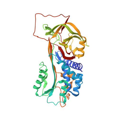
Structures of active and latent PAI-1: a possible stabilizing role for chloride ions.

Stout, T.J.Graham, H.Buckley, D.I.Matthews, D.J.

(2000) Biochemistry 39: 8460-8469

  • DOI: https://doi.org/10.1021/bi000290w
  • Primary Citation of Related Structures:  
    1DVM, 1DVN

  • PubMed Abstract: 

    Serpins exhibit a range of physiological roles and can contribute to certain disease states dependent on their various conformations. Understanding the mechanisms of the large-scale conformational reorganizations of serpins may lead to a better understanding of their roles in various cardiovascular diseases. We have studied the serpin, plasminogen activator inhibitor 1 (PAI-1), in both the active and the latent state and found that anionic halide ions may play a role in the active-to-latent structural transition. Crystallographic analysis of a stable mutant form of active PAI-1 identified an anion-binding site between the central beta-sheet and a small surface domain. A chloride ion was modeled in this site, and its identity was confirmed by soaking crystals in a bromide-containing solution and calculating a crystallographic difference map. The anion thus located forms a 4-fold ligated linchpin that tethers the surface domain to the central beta-sheet into which the reactive center loop must insert during the active-to-latent transition. Timecourse experiments measuring active PAI-1 stability in the presence of various halide ions showed a clear trend for stabilization of the active form with F(-) > Cl(-) > Br(-) >> I(-). We propose that the "stickiness" of this pin (i.e., the electronegativity of the anion) contributes to the energetics of the active-to-latent transition in the PAI-1 serpin.


  • Organizational Affiliation

    MetaXen, South San Francisco, CA 94080, USA. matthews@exelixis.com


Macromolecules
Find similar proteins by:  (by identity cutoff)  |  3D Structure
Entity ID: 1
MoleculeChains Sequence LengthOrganismDetailsImage
PLASMINOGEN ACTIVATOR INHIBITOR-1
A, B, C, D
379Homo sapiensMutation(s): 4 
Gene Names: UMBILICAL VEIN ENDOTHELIUM LIBRARY
UniProt & NIH Common Fund Data Resources
Find proteins for P05121 (Homo sapiens)
Explore P05121 
Go to UniProtKB:  P05121
PHAROS:  P05121
GTEx:  ENSG00000106366 
Entity Groups  
Sequence Clusters30% Identity50% Identity70% Identity90% Identity95% Identity100% Identity
UniProt GroupP05121
Sequence Annotations
Expand
  • Reference Sequence
Experimental Data & Validation

Experimental Data

  • Method: X-RAY DIFFRACTION
  • Resolution: 2.40 Å
  • R-Value Free: 0.292 
  • R-Value Work: 0.218 
  • R-Value Observed: 0.218 
  • Space Group: P 1
Unit Cell:
Length ( Å )Angle ( ˚ )
a = 64.764α = 85.19
b = 74.674β = 86.17
c = 103.445γ = 64.34
Software Package:
Software NamePurpose
X-PLORmodel building
X-PLORrefinement
SCALEPACKdata scaling
X-PLORphasing

Structure Validation

View Full Validation Report


Entry History 

Deposition Data

Revision History  (Full details and data files)

  • Version 1.0: 2000-09-06
    Type: Initial release
  • Version 1.1: 2008-05-05
    Changes: Version format compliance
  • Version 1.2: 2011-07-13
    Changes: Version format compliance
  • Version 1.3: 2017-10-04
    Changes: Advisory, Refinement description
  • Version 1.4: 2021-11-03
    Changes: Advisory, Database references, Derived calculations
  • Version 1.5: 2024-02-07
    Changes: Data collection