Crystal structures of Nova-1 and Nova-2 K-homology RNA-binding domains.
Lewis, H.A., Chen, H., Edo, C., Buckanovich, R.J., Yang, Y.Y., Musunuru, K., Zhong, R., Darnell, R.B., Burley, S.K.(1999) Structure 7: 191-203
- PubMed: 10368286
- DOI: https://doi.org/10.1016/S0969-2126(99)80025-2
- Primary Citation of Related Structures:
1DT4, 1DTJ - PubMed Abstract:
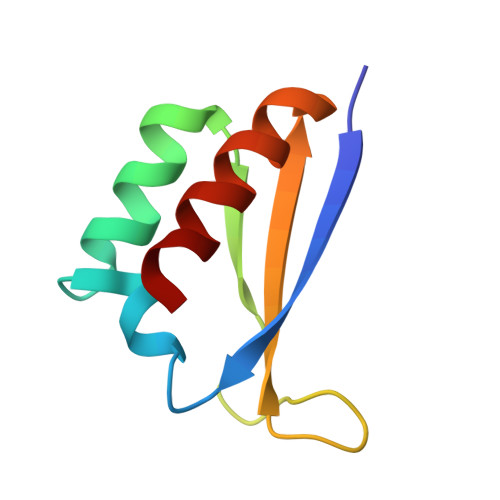
Nova-1 and Nova-2 are related neuronal proteins that were initially cloned using antisera obtained from patients with the autoimmune neurological disease paraneoplastic opsoclonus-myoclonus ataxia (POMA). Both of these disease gene products contain three RNA-binding motifs known as K-homology or KH domains, and their RNA ligands have been identified via binding-site selection experiments. The KH motif structure has been determined previously using NMR spectroscopy, but not using X-ray crystallography. Many proteins contain more than one KH domain, yet there is no published structural information regarding the behavior of such multimers. We have obtained the first X-ray crystallographic structures of KH-domain-containing proteins. Structures of the third KH domains (KH3) of Nova-1 and Nova-2 were determined by multiple isomorphous replacement and molecular replacement at 2.6 A and 2.0 A, respectively. These highly similar RNA-binding motifs form a compact protease-resistant domain resembling an open-faced sandwich, consisting of a three-stranded antiparallel beta sheet topped by three alpha helices. In both Nova crystals, the lattice is composed of symmetric tetramers of KH3 domains that are created by two dimer interfaces. The crystal structures of both Nova KH3 domains are similar to the previously determined NMR structures. The most significant differences among the KH domains involve changes in the positioning of one or more of the alpha helices with respect to the betasheet, particularly in the NMR structure of the KH1 domain of the Fragile X disease protein FMR-1. Loop regions in the KH domains are clearly visible in the crystal structure, unlike the NMR structures, revealing the conformation of the invariant Gly-X-X-Gly segment that is thought to participate in RNA-binding and of the variable region. The tetrameric arrangements of the Nova KH3 domains provide insights into how KH domains may interact with each other in proteins containing multiple KH motifs.
Organizational Affiliation:
Laboratories of Molecular Biophysics, Howard Hughes Medical Institute, The Rockefeller University, 1230 York Avenue, New York, NY, 10021 USA.