The high-resolution structure of the NADP(H)-binding component (dIII) of proton-translocating transhydrogenase from human heart mitochondria.
White, S.A., Peake, S.J., McSweeney, S., Leonard, G., Cotton, N.P., Jackson, J.B.(2000) Structure 8: 1-12
- PubMed: 10673423
- DOI: https://doi.org/10.1016/s0969-2126(00)00075-7
- Primary Citation of Related Structures:
1DJL - PubMed Abstract:
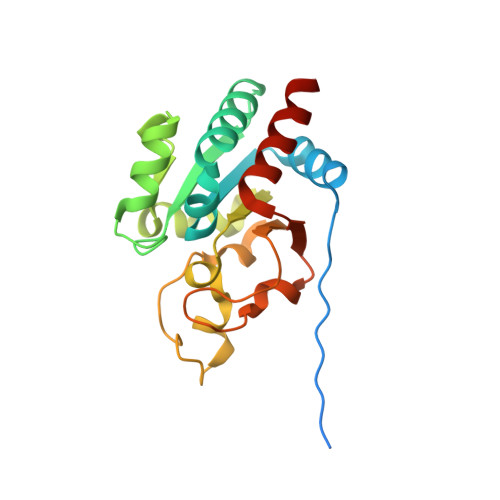
Transhydrogenase, located in the inner membranes of animal mitochondria and the cytoplasmic membranes of bacteria, couples the transfer of reducing equivalents between NAD(H) and NADP(H) to proton pumping. The protein comprises three subunits termed dI, dII and dIII. The dII component spans the membrane. The dI component, which contains the binding site for NAD(+)/NADH, and the dIII component, which has the binding site for NADP(+)/NADPH, protrude from the membrane. Proton pumping is probably coupled to changes in the binding affinities of dIII for NADP(+) and NADPH. The first X-ray structure of the NADP(H)-binding component, dIII, of human heart transhydrogenase is described here at 2.0 A resolution. It comprises a single domain resembling the classical Rossmann fold, but NADP(+) binds to dIII with a reversed orientation. The first betaalphabetaalphabeta motif of dIII contains a Gly-X-Gly-X-X-Ala/Val 'fingerprint', but it has a different function to that in the classical Rossmann structure. The nicotinamide ring of NADP(+) is located on a ridge where it is exposed to interaction with NADH on the dI subunit. Two distinctive features of the dIII structure are helix D/loop D, which projects from the beta sheet, and loop E, which forms a 'lid' over the bound NADP(+). Helix D/loop D interacts with the bound nucleotide and loop E, and probably interacts with the membrane-spanning dII. Changes in ionisation and conformation in helix D/loop D, resulting from proton translocation through dII, are thought to be responsible for the changes in affinity of dIII for NADP(+) and NADPH that drive the reaction.
Organizational Affiliation:
School of Biosciences, University of Birmingham, Edgbaston, B15 2TT, UK. s.a.white@bham.ac.uk