1CY5

CRYSTAL STRUCTURE OF THE APAF-1 CARD


Experimental Data Snapshot

  • Method: X-RAY DIFFRACTION
  • Resolution: 1.30 Å
  • R-Value Free: 0.199 
  • R-Value Observed: 0.169 

wwPDB Validation   3D Report Full Report


This is version 1.3 of the entry. See complete history


Literature

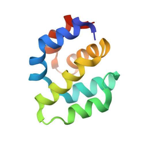
Crystal structure of Apaf-1 caspase recruitment domain: an alpha-helical Greek key fold for apoptotic signaling.

Vaughn, D.E.Rodriguez, J.Lazebnik, Y.Joshua-Tor, L.

(1999) J Mol Biol 293: 439-447

  • DOI: https://doi.org/10.1006/jmbi.1999.3177
  • Primary Citation of Related Structures:  
    1CY5

  • PubMed Abstract: 

    The caspase recruitment domain (CARD) of Apaf-1 binds to the CARD of caspase-9 to trigger a proteolytic cascade that leads to apoptotic cell death. We report the crystal structure of the Apaf-1 CARD at 1. 3 A resolution, solved in a two-element multiwavelength anomalous dispersion (MAD) X-ray diffraction experiment. This CARD adopts a six-helix bundle fold with Greek key topology surrounding an extensive hydrophobic core. This fold, which we call the "death fold", is found in other domains that mediate interactions in apoptotic signaling despite very low sequence identity. From a structure-based alignment, we identify conserved patterns that characterize the death fold and its subclasses. Like the Ig-fold, it provides a rigid structural scaffold upon which diverse recognition surfaces are assembled.


  • Organizational Affiliation

    W. M. Keck Structural Biology, Cold Spring Harbor, NY 11724, USA.


Macromolecules
Find similar proteins by:  (by identity cutoff)  |  3D Structure
Entity ID: 1
MoleculeChains Sequence LengthOrganismDetailsImage
PROTEIN (APOPTOTIC PROTEASE ACTIVATING FACTOR 1)97Homo sapiensMutation(s): 0 
UniProt & NIH Common Fund Data Resources
Find proteins for O14727 (Homo sapiens)
Explore O14727 
Go to UniProtKB:  O14727
PHAROS:  O14727
GTEx:  ENSG00000120868 
Entity Groups  
Sequence Clusters30% Identity50% Identity70% Identity90% Identity95% Identity100% Identity
UniProt GroupO14727
Sequence Annotations
Expand
  • Reference Sequence
Experimental Data & Validation

Experimental Data

  • Method: X-RAY DIFFRACTION
  • Resolution: 1.30 Å
  • R-Value Free: 0.199 
  • R-Value Observed: 0.169 
  • Space Group: P 21 21 21
Unit Cell:
Length ( Å )Angle ( ˚ )
a = 36.35α = 90
b = 46.91β = 90
c = 54.35γ = 90
Software Package:
Software NamePurpose
SHARPphasing
SHELXL-97refinement
DENZOdata reduction
SCALEPACKdata scaling

Structure Validation

View Full Validation Report


Entry History 

Deposition Data

Revision History  (Full details and data files)

  • Version 1.0: 1999-09-13
    Type: Initial release
  • Version 1.1: 2008-04-27
    Changes: Version format compliance
  • Version 1.2: 2011-07-13
    Changes: Version format compliance
  • Version 1.3: 2018-01-31
    Changes: Experimental preparation