1CB2

CELLOBIOHYDROLASE II, CATALYTIC DOMAIN, MUTANT Y169F


Experimental Data Snapshot

  • Method: X-RAY DIFFRACTION
  • Resolution: 2.00 Å
  • R-Value Free: 0.232 
  • R-Value Work: 0.210 
  • R-Value Observed: 0.210 

wwPDB Validation   3D Report Full Report


Ligand Structure Quality Assessment 


This is version 1.4 of the entry. See complete history


Literature

The active site of Trichoderma reesei cellobiohydrolase II: the role of tyrosine 169.

Koivula, A.Reinikainen, T.Ruohonen, L.Valkeajarvi, A.Claeyssens, M.Teleman, O.Kleywegt, G.J.Szardenings, M.Rouvinen, J.Jones, T.A.Teeri, T.T.

(1996) Protein Eng 9: 691-699

  • DOI: https://doi.org/10.1093/protein/9.8.691
  • Primary Citation of Related Structures:  
    1CB2

  • PubMed Abstract: 

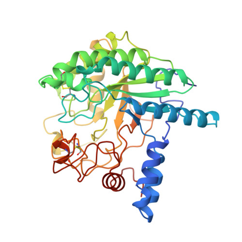
    Trichoderma reesei cellobiohydrolase II (CBHII) is an exoglucanase cleaving primarily cellobiose units from the non-reducing end of cellulose chains. The beta-1,4 glycosidic bond is cleaved by acid catalysis with an aspartic acid, D221, as the likely proton donor, and another aspartate, D175, probably ensuring its protonation and stabilizing charged reaction intermediates. The catalytic base has not yet been identified experimentally. The refined crystal structure of CBHII also shows a tyrosine residue, Y169, located close enough to the scissile bond to be involved in catalysis. The role of this residue has been studied by introducing a mutation Y169F, and analysing the kinetic and binding behavior of the mutated CBHII. The crystal structure of the mutated enzyme was determined to 2.0 A resolution showing no changes when compared with the structure of native CBHII. However, the association constants of the mutant enzyme for cellobiose and cellotriose are increased threefold and for 4-methylumbelliferyl cellobioside over 50-fold. The catalytic constants towards cellotriose and cellotetraose are four times lower for the mutant. These data suggest that Y169, on interacting with a glucose ring entering the second subsite in a narrow tunnel, helps to distort the glucose ring into a more reactive conformation. In addition, a change in the pH activity profile was observed. This indicates that Y169 may have a second role in the catalysis, namely to affect the protonation state of the active site carboxylates, D175 and D221.


  • Organizational Affiliation

    VTT Biotechnology and Food Research, Espoo, Finland.


Macromolecules
Find similar proteins by:  (by identity cutoff)  |  3D Structure
Entity ID: 1
MoleculeChains Sequence LengthOrganismDetailsImage
CELLOBIOHYDROLASE II
A, B
365Trichoderma reeseiMutation(s): 1 
Gene Names: CBH2 (Y169F)
EC: 3.2.1.91
UniProt
Find proteins for P07987 (Hypocrea jecorina)
Explore P07987 
Go to UniProtKB:  P07987
Entity Groups  
Sequence Clusters30% Identity50% Identity70% Identity90% Identity95% Identity100% Identity
UniProt GroupP07987
Sequence Annotations
Expand
  • Reference Sequence
Small Molecules
Ligands 2 Unique
IDChains Name / Formula / InChI Key2D Diagram3D Interactions
NAG
Query on NAG

Download Ideal Coordinates CCD File 
C [auth A],
D [auth A],
L [auth B],
M [auth B]
2-acetamido-2-deoxy-beta-D-glucopyranose
C8 H15 N O6
OVRNDRQMDRJTHS-FMDGEEDCSA-N
MAN
Query on MAN

Download Ideal Coordinates CCD File 
E [auth A]
F [auth A]
G [auth A]
H [auth A]
I [auth A]
E [auth A],
F [auth A],
G [auth A],
H [auth A],
I [auth A],
J [auth A],
K [auth A],
N [auth B],
O [auth B],
P [auth B],
Q [auth B],
R [auth B],
S [auth B],
T [auth B]
alpha-D-mannopyranose
C6 H12 O6
WQZGKKKJIJFFOK-PQMKYFCFSA-N
Experimental Data & Validation

Experimental Data

  • Method: X-RAY DIFFRACTION
  • Resolution: 2.00 Å
  • R-Value Free: 0.232 
  • R-Value Work: 0.210 
  • R-Value Observed: 0.210 
  • Space Group: P 1 21 1
Unit Cell:
Length ( Å )Angle ( ˚ )
a = 49.1α = 90
b = 75.8β = 103.2
c = 92.9γ = 90
Software Package:
Software NamePurpose
HAMLINdata collection
X-PLORrefinement
HAMLINdata reduction

Structure Validation

View Full Validation Report


Ligand Structure Quality Assessment 


Entry History 

Deposition Data

Revision History  (Full details and data files)

  • Version 1.0: 1996-10-14
    Type: Initial release
  • Version 1.1: 2008-03-24
    Changes: Version format compliance
  • Version 1.2: 2011-07-13
    Changes: Non-polymer description, Version format compliance
  • Version 1.3: 2020-07-29
    Type: Remediation
    Reason: Carbohydrate remediation
    Changes: Data collection, Derived calculations, Other, Structure summary
  • Version 1.4: 2021-11-03
    Changes: Database references, Structure summary