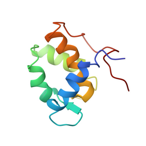
Solution structure of Eps15's third EH domain reveals coincident Phe-Trp and Asn-Pro-Phe binding sites.
Enmon, J.L., de Beer, T., Overduin, M.(2000) Biochemistry 39: 4309-4319
- PubMed: 10757979
- DOI: https://doi.org/10.1021/bi9927383
- Primary Citation of Related Structures:
1C07 - PubMed Abstract:
Eps15 homology (EH) domains interact with proteins involved in endocytosis and signal transduction. EH domains bind to Asn-Pro-Phe (NPF) consensus motifs of target proteins. A few EH domains, such as the third EH domain (EH(3)) of human Eps15, prefer to bind Phe-Trp (FW) sequences. The structure of EH(3) has been solved by nuclear magnetic resonance (NMR) spectroscopy and is the first of an FW- and NPF-binding EH domain. Both FW and NPF sequences bind in the same hydrophobic pocket as shown by heteronuclear chemical shift mapping. EH(3) contains the dual EF-hand fold characteristic of the EH domain family, but it binds calcium with high affinity in the first EF-hand rather than the usual coordination in the second EF-hand. Point mutations were designed based on differences in the EH(3) and the second EH domain (EH(2)) of human Eps15 that alter the affinity of the domains for FW or NPF motif peptides. Peptides that mimic binding sites in the potential EH(3) targets Rab, synaptojanin, and the cation-dependent mannose 6-phosphate receptor were used to explore wild-type and mutant affinities. Characterization of the structure and binding properties of an FW- and NPF-binding EH domain and comparison to an NPF-specific EH domain provide important insights into the mechanisms of EH domain ligand recognition.
Organizational Affiliation:
Department of Chemistry and Biochemistry, University of Colorado, Boulder, Colorado 80309, USA.