X-ray structure of the 154-amino-acid form of recombinant human basic fibroblast growth factor. comparison with the truncated 146-amino-acid form.
Kastrup, J.S., Eriksson, E.S., Dalboge, H., Flodgaard, H.(1997) Acta Crystallogr D Biol Crystallogr 53: 160-168
- PubMed: 15299950
- DOI: https://doi.org/10.1107/S0907444996012711
- Primary Citation of Related Structures:
1BFF - PubMed Abstract:
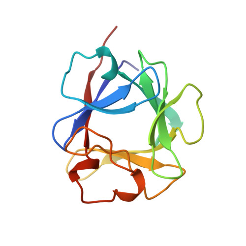
The crystal structure of the 154-amino-acid form of human basic fibroblast growth factor (hbFGF154), probably representing the intact form of hbFGF as deduced from the open reading frame of hbFGF cDNA, was determined by X-ray crystallography and refined to a crystallographic residual of 19.0% for all data between 20.0 and 2.0 A resolution. Crystals were obtained from recombinant hbFGF154 expressed in E. coli. hbFGF154 has the same overall structure as the N-terminus truncated 146-amino-acid form. The structure has a Kunitz-type fold and is built of 12 beta-strands of which six antiparallel strands form a beta-sheet barrel. In the structure it was possible to locate two additional residues at the N terminus and the last three C-terminal amino-acid residues, which seem to be disordered in all but one of the reported structures of the truncated form of hbFGF. The C-terminal amino-acid residues are part of the last beta-strand through the formation of a hydrogen bond between the main-chain amide group of Ala152 and the carbonyl O atom of Pro28. An apparent phosphate ion is bound within the basic region on the surface of the molecule and has as ligands the side chains of Asn35, Arg128 and Lys133 and two water molecules. A slightly different hydrogen-bonding pattern to the phosphate ion is observed as compared with the sulfate ions in the truncated forms [Eriksson, Cousens & Matthews (1993). Protein Sci. 2, 1274-1284; Zhang, Cousens, Barr & Sprang (1991). Proc. Natl Acad. Sci. USA, 88, 3446-3450]. One molecule of beta-mercaptoethanol forms a disulfide bridge to Cys77.
Organizational Affiliation:
PharmaBiotec Research Center, Department of Medicinal Chemistry, Royal Danish School of Pharmacy, Copenhagen, Denmark.