1AYO

RECEPTOR BINDING DOMAIN OF BOVINE ALPHA-2-MACROGLOBULIN


Experimental Data Snapshot

  • Method: X-RAY DIFFRACTION
  • Resolution: 1.90 Å
  • R-Value Free: 0.239 
  • R-Value Observed: 0.195 

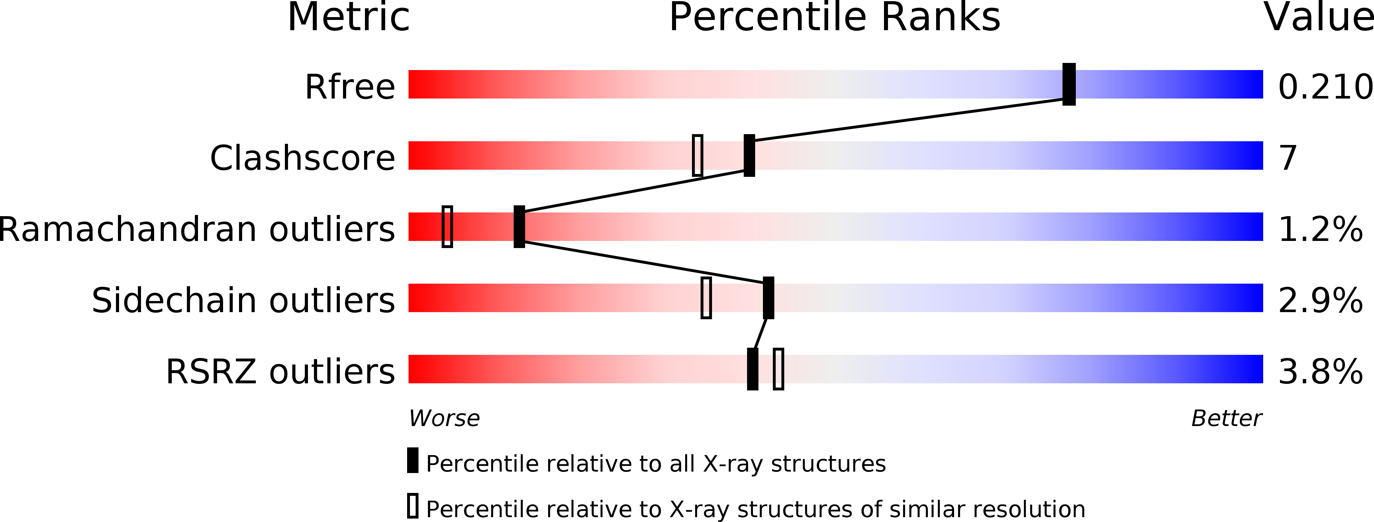
wwPDB Validation   3D Report Full Report


This is version 2.0 of the entry. See complete history


Literature

Crystal structure of the receptor-binding domain of alpha 2-macroglobulin.

Jenner, L.Husted, L.Thirup, S.Sottrup-Jensen, L.Nyborg, J.

(1998) Structure 6: 595-604

  • DOI: https://doi.org/10.1016/s0969-2126(98)00061-6
  • Primary Citation of Related Structures:  
    1AYO

  • PubMed Abstract: 

    The large plasma proteinase inhibitors of the alpha 2-macroglobulin superfamily inhibit proteinases by capturing them within a central cavity of the inhibitor molecule. After reaction with the proteinase, the alpha-macroglobulin-proteinase complex binds to the alpha-macroglobulin receptor, present in the liver and other tissues, and becomes endocytosed and rapidly removed from the circulation. The complex binds to the receptor via recognition sites located on a separate domain of approximately 138 residues positioned at the C terminus of the alpha-macroglobulin subunit.


  • Organizational Affiliation

    Department of Molecular and Structural Biology, University of Aarhus, Denmark.


Macromolecules
Find similar proteins by:  (by identity cutoff)  |  3D Structure
Entity ID: 1
MoleculeChains Sequence LengthOrganismDetailsImage
ALPHA-2-MACROGLOBULIN
A, B
130Bos taurusMutation(s): 0 
UniProt
Find proteins for Q7SIH1 (Bos taurus)
Explore Q7SIH1 
Go to UniProtKB:  Q7SIH1
Entity Groups  
Sequence Clusters30% Identity50% Identity70% Identity90% Identity95% Identity100% Identity
UniProt GroupQ7SIH1
Sequence Annotations
Expand
  • Reference Sequence
Oligosaccharides

Help

Entity ID: 2
MoleculeChains Length2D Diagram Glycosylation3D Interactions
2-acetamido-2-deoxy-beta-D-glucopyranose-(1-4)-2-acetamido-2-deoxy-beta-D-glucopyranose
C
2N-Glycosylation
Glycosylation Resources
GlyTouCan:  G42666HT
GlyCosmos:  G42666HT
GlyGen:  G42666HT
Small Molecules
Ligands 1 Unique
IDChains Name / Formula / InChI Key2D Diagram3D Interactions
CA
Query on CA

Download Ideal Coordinates CCD File 
D [auth B]CALCIUM ION
Ca
BHPQYMZQTOCNFJ-UHFFFAOYSA-N
Experimental Data & Validation

Experimental Data

  • Method: X-RAY DIFFRACTION
  • Resolution: 1.90 Å
  • R-Value Free: 0.239 
  • R-Value Observed: 0.195 
  • Space Group: P 31 2 1
Unit Cell:
Length ( Å )Angle ( ˚ )
a = 107.57α = 90
b = 107.57β = 90
c = 72.229γ = 120
Software Package:
Software NamePurpose
DENZOdata reduction
SCALEPACKdata scaling
SHELXL-97model building
SHELXL-97refinement
SHELXL-97phasing

Structure Validation

View Full Validation Report


Entry History 

Deposition Data

Revision History  (Full details and data files)

  • Version 1.0: 1998-11-25
    Type: Initial release
  • Version 1.1: 2008-03-03
    Changes: Version format compliance
  • Version 1.2: 2011-07-13
    Changes: Non-polymer description, Version format compliance
  • Version 2.0: 2020-07-29
    Type: Remediation
    Reason: Carbohydrate remediation
    Changes: Atomic model, Data collection, Database references, Derived calculations, Structure summary