Crystal structure of ferrochelatase: the terminal enzyme in heme biosynthesis.
Al-Karadaghi, S., Hansson, M., Nikonov, S., Jonsson, B., Hederstedt, L.(1997) Structure 5: 1501-1510
- PubMed: 9384565
- DOI: https://doi.org/10.1016/s0969-2126(97)00299-2
- Primary Citation of Related Structures:
1AK1 - PubMed Abstract:
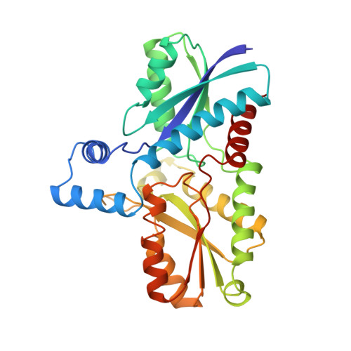
The metallation of closed ring tetrapyrroles resulting in the formation of hemes, chlorophylls and vitamin B12 is catalyzed by specific enzymes called chelatases. Ferrochelatase catalyzes the terminal step in heme biosynthesis by inserting ferrous ion into protoporphyrin IX by a mechanism that is poorly understood. Mutations in the human gene for ferrochelatase can result in the disease erythropoietic protoporphyria, and a further understanding of the mechanism of this enzyme is therefore of clinical interest. No three-dimensional structure of a tetrapyrrole metallation enzyme has been available until now. The three-dimensional structure of Bacillus subtilis ferrochelatase has been determined at 1.9 A resolution by the method of multiple isomorphous replacement. The structural model contains 308 of the 310 amino acid residues of the protein and 198 solvent molecules. The polypeptide is folded into two similar domains each with a four-stranded parallel beta sheet flanked by alpha helices. Structural elements from both domains build up a cleft, which contains several amino acid residues that are invariant in ferrochelatases from different organisms. In crystals soaked with gold and cadmium salt solutions, the metal ion was found to be coordinated to the conserved residue His 183, which is located in the cleft. This histidine residue has previously been suggested to be involved in ferrous ion binding. Ferrochelatase seems to have a structurally conserved core region that is common to the enzyme from bacteria, plants and mammals. We propose that porphyrin binds in the identified cleft; this cleft also includes the metal-binding site of the enzyme. It is likely that the structure of the cleft region will have different conformations upon substrate binding and release.
Organizational Affiliation:
Department of Molecular Biophysics, Lund University, Box 124, S-221 00, Lund, Sweden. salam@hekla.mbfys.lu.se