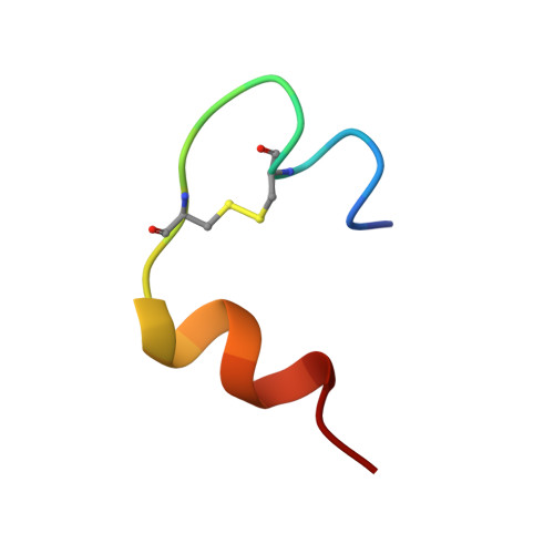
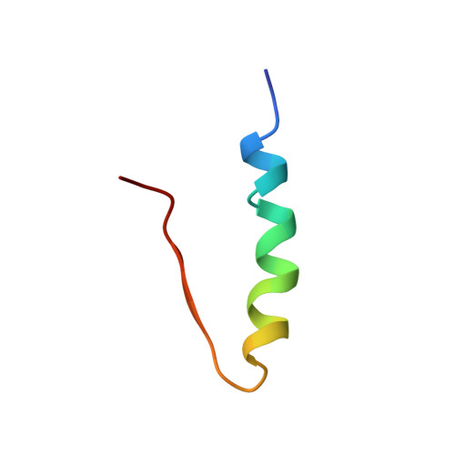
Solution structures of the R6 human insulin hexamer.
Chang, X., Jorgensen, A.M., Bardrum, P., Led, J.J.(1997) Biochemistry 36: 9409-9422
- PubMed: 9235985
- DOI: https://doi.org/10.1021/bi9631069
- Primary Citation of Related Structures:
1AI0, 1AIY - PubMed Abstract:
The three-dimensional solution structure of the phenol-stabilized 36 kDa R6 insulin hexamer was determined by NMR spectroscopy and restrained molecular dynamics. The hexamer structures were derived using a stepwise procedure. Initially, 60 monomers were obtained by distance geometry from 665 NOE-derived distance restraints and three disulfide bridges. Subsequently, the hexamer structures were calculated by simulated annealing, using 30 hexamers constructed from the best 36 monomer structures as the starting models. The NMR data show that the aromatic ring of residue Phe(B25) can take two different orientations in the solution hexamer: one in which it points inward (molecule 1, about 90%) and one in which it points outward from the surface of the monomer (molecule 2, about 10%). Therefore, two hexamer structures were calculated: a symmetric hexamer consisting of six molecule 1 monomers and a nonsymmetric hexamer consisting of five molecule 1 monomers and one molecule 2 monomer. For each of the six monomers, the restraints used in the calculations of the hexamer structures include, in addition to the intramonomeric restraints, 25 NOEs between insulin and phenol, 23 NOEs and two hydrogen bonds across the dimer interface, nine NOEs across the trimer interface, and five intramonomeric or two intermonomeric NOEs, respectively, specifying the different orientations of the Phe(B25) ring. The coordination of the two Zn atoms was defined by eight distance restraints. Thus, a total of 4394 and 4391 distance restraints, respectively, were used in the two hexamer calculations. The NOE restraints were classified in an iterative process as intra- or intermonomeric on the basis of their consistency or inconsistency with the structure of the monomer. The assignment of the dimer- and trimer-specific NOEs was made using the crystal structure of the R6 hexamer as the starting model. For both solution hexamers, the average backbone rms deviation is 0.81 A, if the less well-defined N- and C-terminal residues are excluded. The corresponding rms deviations for all heavy atoms are 1.17 and 1.19 A for the nonsymmetric and symmetric hexamer, respectively. The overall solution structure of the R6 insulin hexamer is compact, rigid, and symmetric and resembles the corresponding crystal structure. However, the extension of the B-chain alpha-helix, which characterizes the R state, is shorter in the solution structure than in the crystal structure. Also, the study shows that the orientation of the Phe(B25) ring has no effect on the structure of the rest of the molecule, within the uncertainty of the structure determination. The importance of these findings for the current model for the insulin-receptor interaction is discussed.
Organizational Affiliation:
Department of Chemistry, University of Copenhagen, The H. C. Orsted Institute, Universitetsparken 5, DK-2100 Copenhagen O, Denmark.