The structure of the Escherichia coli phosphotransferase IIAmannitol reveals a novel fold with two conformations of the active site.
van Montfort, R.L., Pijning, T., Kalk, K.H., Hangyi, I., Kouwijzer, M.L., Robillard, G.T., Dijkstra, B.W.(1998) Structure 6: 377-388
- PubMed: 9551558
- DOI: https://doi.org/10.1016/s0969-2126(98)00039-2
- Primary Citation of Related Structures:
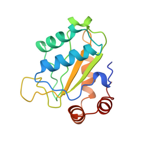
1A3A - PubMed Abstract:
The bacterial phosphoenolpyruvate-dependent phosphotransferase system (PTS) catalyses the cellular uptake and subsequent phosphorylation of carbohydrates. Moreover, the PTS plays a crucial role in the global regulation of various metabolic pathways. The PTS consists of two general proteins, enzyme I and the histidine-containing protein (HPr), and the carbohydrate-specific enzyme II (EII). EIIs are usually composed of two cytoplasmic domains, IIA and IIB, and a transmembrane domain, IIC. The IIA domains catalyse the transfer of a phosphoryl group from HPr to IIB, which phosphorylates the transported carbohydrate. Knowledge of the structures of the IIA proteins may provide insight into the mechanisms by which the PTS couples phosphorylation reactions with carbohydrate specificity. We have determined the crystal structure of the Escherichia coli mannitol-specific IIA domain, IIAmtl (M(r) 16.3 kDa), by multiple anomalous dispersion analysis of a selenomethionine variant of IIAmtl. The structure was refined at 1.8 A resolution to an R factor of 19.0% (Rfree 24.2%). The enzyme consists of a single five-stranded mixed beta sheet, flanked by helices on both sides. The phosphorylation site (His65) is located at the end of the third beta strand, in a shallow crevice lined with hydrophobic residues. The sidechains of two conserved active-site residues, Arg49 and His111, adopt two different conformations in the four independent IIAmtl molecules. Using a solution structure of phosphorylated HPr, and a combination of molecular modelling and NMR binding experiments, structural models of the HPr-IIAmtl complex were generated. The fold of IIAmtl is completely different from the structures of other IIA proteins determined so far. The two conformations of Arg49 and His111 might represent different states of the active site, required for the different phosphoryl transfer reactions in which IIAmtl is involved. A comparison of the HPr-IIAmtl model with models of HPr in complex with other IIA enzymes shows that the overall interaction mode between the two proteins is similar. Differences in the stabilisation of the invariant residue Arg17 of HPr by the different IIA proteins might be part of a subtle mechanism to control the hierarchy of carbohydrate utilisation by the bacterium.
Organizational Affiliation:
Laboratory of Biophysical Chemistry, BIOSON Research Institute, University of Groningen, Nijenborgh, The Netherlands.