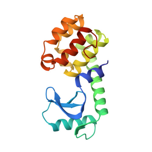
A helix initiation signal in T4 lysozyme identified by polyalanine mutagenesis.
Zhang, X.J., Baase, W.A., Matthews, B.W.(2002) Biophys Chem 101-102: 43-56
- PubMed: 12487988
- DOI: https://doi.org/10.1016/s0301-4622(02)00193-x
- Primary Citation of Related Structures:
190L, 191L, 192L - PubMed Abstract:
To better understand the relation between sequence and structure, and in an attempt to simplify the protein folding problem, a series of alanine substitutions was introduced into bacteriophage T4 lysozyme. In contrast to previous studies in this system, which were restricted to single alpha-helices, the present analysis included a helix-turn-helix region, a loop-helix region, and two alpha-helices that were well separated in the three-dimensional structure. It was shown previously that T4 lysozyme is very tolerant of alanine substitutions within alpha-helices, especially at solvent-exposed sites. The present study shows that the protein is also tolerant of such substitutions in turn and loop regions, although less than in helices. The results confirm that the structural information in the amino acid sequence is highly redundant. For example, the protein with the sequence 127AAAAAALAAAAWAAA141 folds normally, has melting temperature only 0.8 degrees C lower than wildtype, and has a crystal structure that is also very similar to wildtype. Polyalanine substitutions within turns or loops can, however, lead to differences in structure and in folding. In one example the triple substitution K35A/S36A/P37A caused this region of the molecule to change to a more helical conformation. In a second case the mutant with the sequence 34AAAAALAAAKAALAAA49, which spans a loop-helix region, had a dramatically altered thermal unfolding transition, suggesting that this region may tend to form a single, uninterrupted, helix. Substitution of Ala38 in the above construct with aspartic acid caused the unfolding to be more like wildtype, suggesting that residue 38, which is at a helix-capping position in the wildtype structure, provides an initiation signal that is essential in the polyalanine mutant for the correct formation of alpha-helix 39-50. In a typical protein, the information that codes for the 3D structure is presumably distributed over many amino acids. The present results suggest that in simplified sequences the key folding information may be restricted to a subset of critical residues, and so be more readily accessible to experimental analysis.
Organizational Affiliation:
Institute of Molecular Biology, Howard Hughes Medical Institute and Department of Physics, University of Oregon, Eugene, OR 97403-1229, USA.