Rapid crystallization of T4 lysozyme by intermolecular disulfide cross-linking.
Heinz, D.W., Matthews, B.W.(1994) Protein Eng 7: 301-307
- PubMed: 8177878
- DOI: https://doi.org/10.1093/protein/7.3.301
- Primary Citation of Related Structures:
138L, 139L - PubMed Abstract:
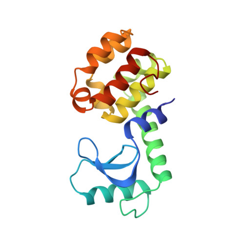
In an attempt to facilitate crystallization, engineered cysteines were used to promote formation of a 'back-to-back' dimer that occurs in different crystal forms of wild-type and mutant T4 lysozymes. The designed double mutant, N68C/A93C, in which the surface residues Asn68 and Ala93 were replaced by cysteines, formed dimers in solution and crystallized isomorphously to wild-type, but at a much faster rate. Overall, the mutant structure remained very similar to wild-type despite the formation of two intermolecular disulfide bridges. The crystals of cross-linked dimers ahd thermal factors somewhat lower than wild-type, indicating reduced mobility, but did not diffract to noticeably higher resolution. Introduction of the same cross-links was also used to obtain crystals in a different space group of a T4 lysozyme mutant that could not be crystallized previously. The results suggest that the formation of the lysozyme dimer is a critical intermediate in the formation of more than one crystal form and that covalent cross-linking of the intermediate accelerates nucleation and facilitates crystal growth. The disulfide cross-links are located on the 'back' of the molecule and formation of the cross-linked dimer appears to leave the active sites completely unobstructed. Nevertheless, the cross-linked dimer is completely inactive. One explanation for this behavior is that the disulfide bridges prevent hinge-bending motion that may be required for catalysis. Another possibility is that the formation of the dimer increases the overall bulk of the enzyme and prevents its access to the susceptible glycosidic bonds within the cell wall substrate.
Organizational Affiliation:
Institute of Molecular Biology, Howard Hughes Medical Institute, Eugene, OR.